


Mediastinum and Hila
Chest X-rays; The radiographic density of the hilum is largely due to the upper lobe veins and pulmonary arteries. The hila should be symmetrical in size and density. If the patient’s posture during the radiograph is not rotated or if there is no abnormality like scoliosis in the spine, a hilum that is larger or denser than the other raises suspicion of hilar pathology.
The most common cause of bilateral hilar enlargement is the enlargement of lymph nodes. Other causes include vascular pathologies and hilar-bronchial masses, which are responsible for the enlargement of these lesions.
Heart and Pulmonary Vessels
In a posteroanterior (PA) radiograph, the right border of the mediastinum is formed by the right atrium below and the ascending aorta above. The superior vena cava is also located in this region, but it is not usually visible unless it is dilated. The left border is made up of the aortic knob at the top, followed by the pulmonary conus, and then the left atrial appendage and left ventricle below.
Pleura and Pericardium
In a standing PA chest radiograph, pleural effusions less than 200 mL may not be seen. Since the posterior costophrenic angles are lower than the lateral costophrenic angles, a lateral chest radiograph may show smaller effusions. The most sensitive position for detecting effusions is a lateral decubitus radiograph, where even an effusion as small as 15-20 mL can be detected. The signs of pleural effusion on a standing PA chest radiograph include the appearance of an elevated hemidiaphragm, blunting of the lateral costophrenic angle, a meniscus sign (a curving line that is concave upwards, higher on the outer edges and lower toward the center), and the mediastinum being pushed to the opposite side in cases of large amounts of fluid.
Detecting pleural effusion on a supine radiograph can be difficult. A veil-like opacity may appear without obscuring vascular markings, and sometimes the costophrenic angle or the lung apex may be obscured. In some cases, a large portion of the pleural effusion accumulates between the lower surface of the lung and the diaphragm (subpulmonic effusion). In this case, the PA chest radiograph may show an elevated, flattened, and laterally displaced hemidiaphragm. Subpulmonic effusion is more common on the right side, but when it occurs on the left, the distance between the gas in the stomach/fundus and the lung base increases to more than 2 cm.
Lungs
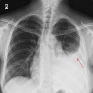
Radiographic findings of atelectasis include increased density of the atelectatic lung parenchyma, displacement of fissures, hilum, and mediastinum, crowding of vessels and bronchi, diaphragm elevation, narrowing of the intercostal spaces, and compensatory hyperinflation of the normal lung.
Airspace disease follows an acinar pattern, where fluid and/or cells replace air in the alveoli or acinar lumen. A nodule is a round or oval opacity, 3 cm or smaller, surrounded by lung or visceral pleura, without associated lymphadenopathy, atelectasis, or pneumonia. Lesions larger than 3 cm are called masses.
Differential diagnosis of solitary nodules and masses typically requires advanced imaging techniques such as PET-CT or MRI.
Chest X-ray Prices 2026
For more information on chest X-ray prices in 2026, feel free to contact us.
——————————————