


Dental Tomography Dental Volumetric Tomography ( 3D VT)
Dental tomography device is a device that converts the data obtained from the jaw and teeth region into a three-dimensional image and transfers it to the digital environment. The resulting images make the diagnosis of diseases even easier.
Dental tomography Dental Volumetric Tomography (3D VT) device is a new tomography device specially prepared for the head and neck region. The desired area can be seen in 3-dimensional tomographically so that the patient is exposed to a very small amount of radiation.
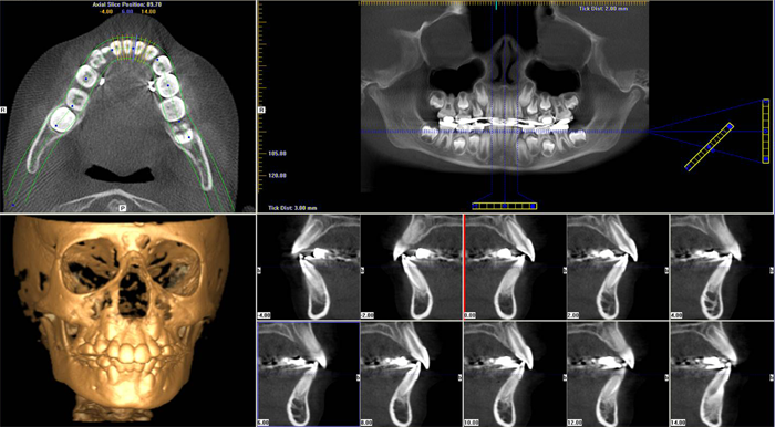
A large number of sections are taken from the axial, sagittal and coronal planes with a dental tomography device. Later, these sections are re-sliced and configured with advanced software so that the targeted area can be viewed from the desired angle and from all directions.
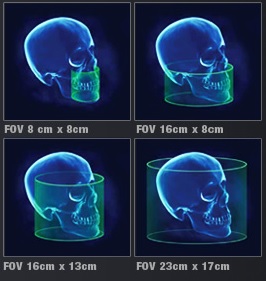
I-CAT 3D dental volumetric tomography device is used in our center. i-CAT holds the title of the device that takes the largest 3-dimensional image with its 3-dimensional and cylindrical image area of 23 (diameter) x17 (height) cm.
When should I have a dental tomography (3D dental volumetric tomography 3d vt ) taken ?
It is often used for dental and maxillofacial diseases, dental implant applications and trauma to the maxillofacial region. It provides detailed diagnosis in dental and maxillofacial pathologies.
The most important place of use of dental tomography ( Dental Volumetric Tomography ) is dental implant applications, which are becoming very common today.
Treatment planning before dentomaxillofacial surgery applications
Preparation of surgical template for implant placement
Anatomical examination of the nasal cavity, incisional canal, maxillary sinus and mandibular canal, such as
Measurement of bone quality and density
Examination of jawbone contours
Examination of the temporomandibular joint
Examination of periapical region and canal fillings in canal treatments
3-dimensional analysis of the positions of embedded teeth in bone
Examination of pathological formations such as cysts and tumors
Examination of root fractures
What are the advantages of dental tomography ( dental volumetric tomography ) ?
As a result of three-dimensional planning with dental volumetric tomography before the placement of dental implants, both possible complications that may occur in the future can be prevented and appropriate response to the aesthetic expectations of patients can be provided.
While only panoramic radiographs were used in implant planning until recently, Dental Volumetric Tomography has also taken an important place in implant planning with the developing technology.
Three-dimensional imaging of the vertical and horizontal amounts of the bone to be implanted allows the physician to start the operation with safer information and thus reduce the undesirable complications that may occur during implant operations.
Shooting takes place with up to 1/6 less radiation than a medical tomography. The amount of radiation given is only 0.5- up to 1.5 panoramic X-rays. The image quality and resolution are very high.
You can view areas that cannot be exposed with panoramic and periapical X-rays. You can take coronal, axial, sagittal (cross section) sections from the taken image and make a more detailed diagnosis.
You can obtain high-resolution data for implant planning and surgical manual preparation. You can make pre-treatment plans such as soft tissue detection, bone density measurement, mandibular canal distance measurement. You can make postoperative checks after treatment, after prosthesis and after implant.You can make determinations such as buried teeth, cracks, abnormal number of channels, cyst spread.
How Much Does It Cost ?
Dr.Abdullah Jawahir
Radiology Specialist
Hekimoglu Imaging and Diagnostic Center