


Transvaginal Color Doppler Ultrasound
Ultrasound is an integral part of modern gynecological examination. Transvaginal ultrasound has allowed for a much more detailed examination of gynecological physiological and pathological processes by evaluating the organs of interest more closely with a high-frequency probe. In addition to the gray-scale views of the ovaries and endometrium, which change throughout the menstrual cycle, the use of Doppler allows for the evaluation of blood flow in these organs. Moreover, most pathological processes result in changes to the normal vascular pattern. It is important to distinguish these changes from the physiological neovascularization that occurs in the ovary around the follicle developing during each menstrual cycle.
Anatomy
Accurate identification of pelvic vessels is essential for proper Doppler evaluation and interpretation. In gynecological applications, emphasis is placed on the uterine and ovarian vessels, but the relationships of other major pelvic arteries and veins are also important. Gynecological masses can compress the iliac veins, and an iliac artery pathology, such as an aneurysm, can mimic an adnexal mass.
After the iliac artery exits the aortic bifurcation, it courses downward and laterally to extend into the lower extremity as the common femoral artery. The approximate surface landmarks for this route are along a line drawn from the navel to the pulse point in the groin. The main and external iliac veins pass behind and medially to their accompanying arteries. These arteries and veins often form the lateral boundary of the ovaries. The internal iliac artery branches medially from the main iliac artery about 4 cm from the aortic bifurcation. The internal iliac artery divides into anterior and posterior branches, and the uterine artery arises from the anterior branch. The uterine artery gives off branches to the upper vagina and cervix as it passes medially to the cervix at the base of the broad ligament. It then ascends within the broad ligament, giving off branches to the myometrium, until it reaches the cornual region, where it turns laterally to supply the fallopian tube and ovary. The uterine vein runs alongside the artery and eventually drains into the internal iliac vein.
The ovarian artery arises from the aorta just below the renal arteries. It travels behind the paracolic gutter in the retroperitoneum, crossing the ureter and psoas muscle. At the pelvic brim, it enters the suspensory ligament of the ovary and extends to the lateral end of the broad ligament beneath the fallopian tube. Its branches supply the ovary and fallopian tube, as well as anastomosing with branches of the uterine artery. As a result, the ovary has dual blood supply. The ovarian veins originate from the venous plexus in the mesovarium and suspensory ligament and accompany the ovarian artery, draining into the renal vein on the left and the inferior vena cava on the right.
How Transvaginal Color Doppler is Performed
Transvaginal scanning allows for a closer evaluation of pelvic vessels, particularly facilitating the visualization of the ovarian artery.
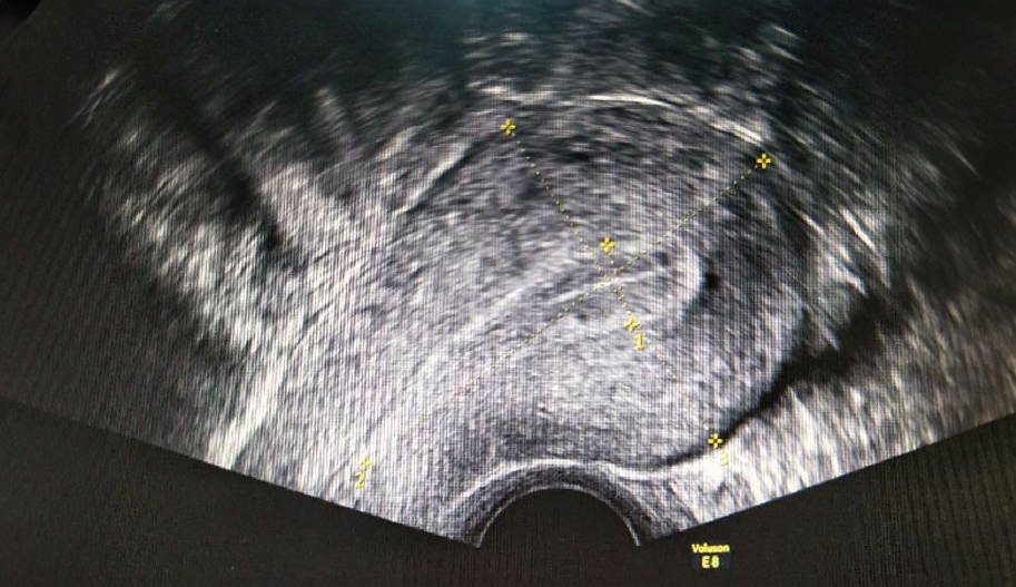
The area of interest is first identified on gray-scale ultrasound. Color Doppler uses a color box, the size of which should be adjusted to fit the size of the area being examined.
Menstrual Disorders
Dysfunctional uterine bleeding is one of the most common gynecological complaints. In most cases, an underlying hormonal cause is identified, but in about half of these cases, ultrasound can reveal an underlying structural abnormality—the most common being submucosal fibroids, adenomyosis, or endometrial polyps. Since women with menstrual pain have been shown to have increased myometrial vascularization early in their menstrual cycle, and those with irregular bleeding are more likely to show increased perfusion in the uterine and subendometrial blood vessels, Doppler plays a more general role in these cases.
Fertility
Normal Cycle
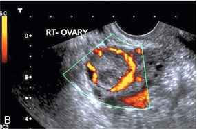
The normal menstrual cycle begins on the first day of bleeding, signaling the shedding of the endometrium. Ultrasound shows the endometrium thinning to the basal layer, and sometimes blood and clots may be present in the cavity. The endometrium then enters the proliferative phase, displaying a characteristic trilaminar appearance. As ovulation approaches, the basal endometrium becomes more echogenic; this echogenicity extends across the entire endometrial thickness during the secretory phase after ovulation. During the proliferative phase, antral follicles in the ovary develop until one becomes dominant. This dominant follicle continues to grow until it reaches about 2.5 cm in diameter, at which point it ruptures, leading to ovulation (usually around day 14). The follicle then forms the corpus luteum during the secretory phase. The increased vascularization around the corpus luteum has been likened to a ring of fire.
Ovarian Reserve
Fertility units, in particular, need to determine ovarian reserve to help assess whether an in vitro fertilization (IVF) procedure will be successful. Typically, an antral follicle count of fewer than four or an ovarian volume <3 cc, measured during the first few days of the menstrual cycle, is associated with a higher likelihood of pregnancy failure or IVF cycle cancellation.
In postmenopausal ovaries, there is typically no detectable blood flow, or there is only high-impedance flow in the ovarian arteries without diastolic flow. The same is true for postmenopausal uterine arteries, where there is also no diastolic flow.
Why Is Transvaginal Doppler Ultrasound Performed?
Doppler evaluation of the pelvis may include, but is not limited to, the following:
– The size, shape, and position of the uterus and ovaries
– The presence of fluid or masses within or near the endometrium, myometrium, fallopian tubes, or bladder
– The length and thickness of the cervix
– Changes in the structure of the bladder
– Blood flow from pelvic organs
Pelvic Doppler can provide a lot of information about the size, location, and structure of pelvic masses, but it cannot provide a definitive diagnosis of cancer or specific diseases. A pelvic Doppler may be used to help diagnose and treat the following conditions:
– Abnormalities in the anatomy of the uterus
– Fibroid tumors (benign growths), masses, cysts, and other types of tumors within the pelvis
– The presence and location of an intrauterine contraceptive device (IUD)
– Pelvic inflammatory disease (PID) and other types of inflammation or infection
– Postmenopausal bleeding
– Monitoring ovarian follicle size for infertility evaluation
– Follicular fluid and egg aspiration from the ovaries for in vitro fertilization
– Ectopic pregnancy (pregnancy occurring outside the uterus, typically in the fallopian tube)
– Monitoring fetal development during pregnancy
– Evaluation of certain fetal conditions
Your doctor may have other reasons for requesting a pelvic Doppler.
Transvaginal Color Doppler Ultrasound Prices 2026
Transvaginal color Doppler ultrasound prices may vary regionally. Even within Istanbul, different neighborhoods may have different prices. For more detailed information about transvaginal color Doppler prices, feel free
to call us at 02126321059 or 05359214295.