


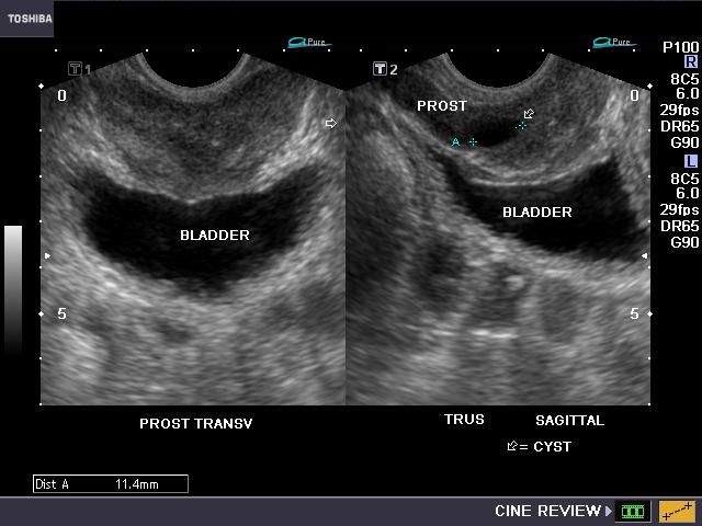
What is Transrectal Ultrasound for Prostate Imaging?
Transrectal ultrasound is a safe and painless procedure that uses sound waves to image the internal parts of the body. In ultrasound imaging, also known as sonography or ultrasound scanning, we use a small transducer (probe) placed directly on the skin and ultrasound gel.
High-frequency sound waves are transmitted from the probe into the body through the gel. The transducer collects the returning sound waves, and a computer converts them into images. Ultrasound examinations do not use ionizing radiation (as is used in X-rays), so the patient is not exposed to radiation.
Since ultrasound images are captured in real-time, they can show the structure and movement of internal organs and blood flowing through blood vessels. Ultrasound imaging is a non-invasive medical test that helps doctors diagnose and treat medical conditions.
Transrectal ultrasound, also known as prostate ultrasound, is an imaging technique used to visualize the prostate gland and surrounding tissues in men. The exam is typically performed by inserting an ultrasound probe into the patient’s rectum. The probe sends and receives sound waves through the rectal wall to the prostate gland, which is located directly in front of the rectum.
What are the uses of transrectal prostate ultrasound?
Transrectal prostate ultrasound is used to detect and evaluate the following:
- Detect abnormalities within the prostate.
- Determine if the prostate is enlarged and if any treatment is necessary in cases of prostate enlargement, also known as benign prostatic hyperplasia (BPH), through measurements taken during the ultrasound.
- Identify any abnormal growths in the prostate.
- Assist in diagnosing the cause of male infertility.
Transrectal ultrasound of the prostate is typically used to help diagnose the following symptoms:
- A nodule felt by the doctor during a routine physical exam or prostate cancer screening.
- Elevated PSA levels in a blood test.
- Difficulty urinating.
Since ultrasound provides real-time images, it can also be used to guide procedures such as needle biopsies, where tissue (cells) are taken from an abnormal area in the prostate for further lab testing.
How is transrectal prostate ultrasound performed?
In men, the prostate gland is located directly in front of the rectum, so transrectal ultrasound is performed to position the ultrasound probe as close to the prostate as possible.
For transrectal ultrasound, you will be asked to lie on your side with your top leg bent at the knee. A disposable protective cover is placed over the probe, and the probe is inserted into the rectum.
Images are obtained from different angles to get the best possible view of the prostate gland.
If a suspicious lesion is detected on ultrasound or during a rectal exam, a biopsy may be performed under ultrasound guidance. During this procedure, the radiologist guides a needle into the prostate gland while monitoring its position on the ultrasound. A small tissue sample is taken for microscopic examination. This shows an example of a transrectal transducer (probe).
A prostate-specific antigen (PSA) test, which measures the amount of PSA in the blood, can be performed to determine whether a patient is at high risk for cancer. If the test result exceeds a certain threshold, a biopsy is performed, and an ultrasound probe is used to guide the biopsy to specific areas of the prostate.
Once the examination is complete, you may be asked to dress and wait while the ultrasound images are reviewed.
This ultrasound examination is usually completed in less than 20 minutes.
Transrectal Ultrasound Prices 2026
For information on transrectal ultrasound prices for 2026, you can contact us immediately.
What is the cost?
Dr. Abdullah Cevahir
Radiology Specialist
Hekimoğlu Imaging and Diagnostic Center