


Transcranial and transfontanel ultrasound, also known as cranial ultrasound, uses sound waves to obtain images of the brain and the ventricles filled with cerebrospinal fluid located in the deeper parts of the brain.
This imaging test is mostly done in infants. It can be used to check the ventricles in babies up to 18 months old and to address issues related to premature birth.
Ultrasound waves cannot pass through bones. Therefore, transcranial ultrasound cannot be performed once the skull bones are fully matured.
Ultrasound is widely available, easy to use, and cheaper than many other imaging methods.
Ultrasound imaging is extremely safe and does not use radiation.
Ultrasound scanning provides clear images of soft tissues, which do not show well on X-rays.
Why is transcranial/transfontanel cranial ultrasound performed for babies?
Periventricular leukomalacia (PVL) is a condition where brain tissue around the ventricles is damaged. This may occur due to lack of oxygen or blood flow before, during, or after birth, causing damage to the brain tissue.
Brain hemorrhage; bleeding into the ventricles and periventricular leukomalacia increases the risk of disability in the baby. This can range from mild learning or motor skill delays to cerebral palsy or intellectual disabilities.
Bleeding into the ventricles is more common in premature babies than in full-term babies. It most often occurs within the first 3 to 4 days after birth.
Most cases of bleeding into the ventricles can be detected by transcranial ultrasound within the first week after birth. However, it may take several weeks to detect PVL. If PVL is suspected, cranial ultrasound can be repeated 4 to 8 weeks after birth.
Cranial ultrasound may be performed if the baby’s head circumference is larger than normal to check for abnormalities. Transcranial ultrasound can also be used to detect infections in or around the brain (such as encephalitis or meningitis) or to check for congenital brain problems (such as congenital hydrocephalus).
Advantages of cranial ultrasound
Accessibility, mobility, bedside scanning in the neonatal ward, no need for sedation,
the ability to enable serial scans, for example, to assess brain maturation and/or lesion development, with no ionizing radiation.
Disadvantages of cranial ultrasound
Like all ultrasound examinations, cranial ultrasound is largely operator-dependent. This is not a real disadvantage but requires the radiographer performing the examination to be well-trained in acquisition techniques, to know how to use the available ultrasound equipment, and to be knowledgeable about intracranial anatomy and pathology.
A natural limitation of cranial ultrasound is that all of the structures forming the acoustic windows will eventually close.
Indications for cranial ultrasound
Routine cranial ultrasound for all premature newborns, suspected brain abnormalities on antenatal ultrasound, any newborn with suspected brain pathology, or a newborn that was not screened before birth.
Cranial Ultrasound Technique
For routine screening, the anterior fontanel serves as an acoustic window. Additional acoustic windows used to image specific intracranial structures include: the posterior fontanel, mastoid fontanel, squamous portion of the temporal bone (i.e., temporal window), foramen magnum, coronal suture, and squamous sutures.
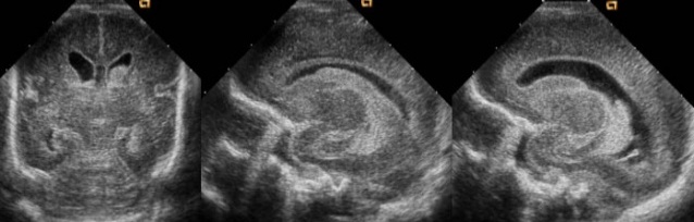
A basic routine screening is performed through the anterior fontanel. To be clear, the brain of a premature newborn (i.e., 28 weeks or less) will appear less developed than that of a term newborn, including less convoluted gyri and shallower sulci; the following should only serve as a rough guide.
In a coronal (transverse) scan, the transducer is first tilted forward, then gradually tilted backward.
Transcranial and Transfontanel Ultrasound Prices 2026
For information on transcranial and transfontanel ultrasound prices for 2026, you can contact us immediately.
Dr. Abdullah Cevahir
Radiology Specialist
Hekimoğlu Imaging and Diagnostic Center