


Spinal sacral ultrasound is the preferred method in newborns suspected of occult spinal dysraphism (OSD).
OSD refers to the presence of one or more spinal cord anomalies that can cause tethering of the spinal cord and potentially lead to neurological, bladder, or bowel dysfunction.
Since the posterior arch of the spine has not yet ossified, providing an excellent acoustic window, ultrasound is easy to perform.
Especially the conus medullaris, cauda equina, and lumbosacral part of the spinal canal can be beautifully visualized with a high-resolution linear probe.
Classification of spinal dysraphism
Spina bifida aperta
Spinal dysraphism, or spina bifida, is a congenital anomaly caused by defective closure of the neural arch. It is classified as open (spina bifida aperta) and closed dysraphism (spina bifida occulta).
Open dysraphism manifests as a swelling on the back noticed at birth, which may contain meninges and CSF, called a meningocele, or spinal cord or nerve parts, called a myelomeningocele.
Ultrasound should not be used to image open spinal dysraphism. It does not provide much information and may lead to infection.
Ultrasound can be useful for examining more cranial parts of the vertebral column, investigating additional anomalies, and measuring the size of the brain’s ventricles after myelomeningocele closure.
Spina bifida occulta
In closed or occult spinal dysraphism, also called spina bifida occulta, the skin remains intact. Anomalies are suspected when skin changes such as tufts of hair, hemangiomas, pigmented spots, cutaneous dimples, or subcutaneous masses are present.
Another reason for performing ultrasound is the presence of a congenital anomaly associated with OSD, such as anal atresia.The term OSD refers to the presence of one or more spinal cord anomalies that can lead to tethering of the spinal cord and potentially cause neurological and bladder/bowel dysfunction.
Thickened or fatty filum terminale, spinal lipomas, split cord malformations, dermoid cysts, and syringohydromyelia are all terms describing different forms of OSD.
Normal anatomy
The spinal cord is depicted as a hypoechoic structure with a central echogenic area.This central echogenicity is expected to represent the interface between the anterior commissure and the median anterior fissure, not the central canal.

Axial image of the spinal cord with central echogenicity.

Axial image of the cauda equina.
The lower end of the cord is thickened by the lumbar enlargement.The cord tapers into a sharp cone (blue arrow).The cauda equina is seen as a group of moving threads.If the baby is lying in a decubitus position, the nerve fibers will be pulled to the back due to gravity.
If the baby is examined prone with a pillow under the abdomen, the nerve fibers will move ventrally.The filum terminale can be seen as a thin echogenic thread.The dural sac ends around S2, with fatty tissue present more distally.
Position of the conus medullaris
The normal position of the conus is at L1. It should not be below L2.The best way to determine the position of the conus medullaris is to identify the lumbosacral junction at the lordotic angle between the lumbar and sacral vertebrae.Flexing and extending the pelvis can help visualize the hinge point of the sacrum.In this newborn, the lumbosacral junction is less distinct because there is no acute angle.
If the coccyx has not yet ossified, it consists of hypoechoic cartilage. It usually has a kyphotic shape and should not be confused with fluid accumulation or an abscess in transverse views.
Normal variants
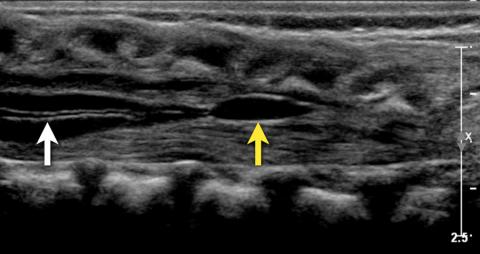
Central canal
In this image, the central canal is seen as a thin anechoic line (white arrow) within the spinal cord.Although sometimes associated with pathology, it is often considered a normal finding.
Ventriculus terminalis
A small cyst is seen in the proximal filum terminale. This is called the terminal ventricle (or fifth ventricle). It is sometimes also seen in the conus medullaris. It forms during embryogenesis and usually regresses completely in early childhood.
If it persists, it is typically less than 2 cm in craniocaudal size and less than 2 mm in transverse size, and is detected as an asymptomatic and incidental finding in adults.

Here is a sagittal image of a three-month-old girl who was scanned due to a skin discoloration on her lower back. The spinal anatomy was normal with no sign of OSD. She has a straight coccyx, which is a normal variant. The coccyx typically has an anteriorly curved tip but can sometimes be straight or even dorsally bent, which is also a normal variant.
Tethered cord
In most cases, occult spinal dysraphism may not cause any symptoms. However, in some cases, neurological issues may develop due to tethering of the cord.
Tethered cord is the pathological fixation of the spinal cord in an abnormally caudal position. In this condition, the spinal cord undergoes mechanical stretching, bending, and ischemia with growth and development.
In such cases, ultrasound is a very suitable imaging method to visualize the contents of the spinal canal and look for findings associated with tethered cord.
Hydromyelia
Hydromyelia is an abnormal dilation of the central canal, filled with cerebrospinal fluid. This condition can occur along the entire length of the spinal cord or in a localized area.
It can be associated with many congenital anomalies, such as diastematomyelia, Arnold-Chiari malformation, myelomeningocele, and lipomeningocele.If fluid accumulation occurs outside the central canal within the cord, it is called syringomyelia.
Since it is often not possible to distinguish between these two conditions on imaging, the term syringohydromyelia is preferred.
Spinal lipoma
A spinal lipoma is a mass of encapsulated fat, nerve tissue, meninges, or fibrotic tissue extending from a midline defect in the fascia into the posterior subcutaneous tissue. A spinal lipoma is usually seen as an echogenic mass in the lower spinal canal. The conus may be very low and embedded within the lipoma.
Dorsal dermal sinus
A connection between the skin and the dural sac, sometimes extending to the spinal cord. It presents with skin discoloration or hairy patches. The conus medullaris may be positioned lower than normal.
Dermal sinus tracts are particularly important lesions to recognize because they can extend into the spinal canal and increase the risk of infections such as meningitis or spinal abscess.
Diastematomyelia
Diastematomyelia, also known as split cord malformation, is a longitudinal splitting of the spinal cord. A bony, fibrous, or cartilaginous septum may be present at the point of division.
Diastematomyelia is usually associated with a vertebral column anomaly and tethered cord.
Spinal/Sacral Ultrasound Prices 2026
For information on spinal/sacral ultrasound prices for 2026, you can contact us immediately.
What is the cost?
Dr. Abdullah Cevahir
Radiology Specialist
Hekimoğlu Imaging and Diagnostic Center