


What is Obstetric Doppler Ultrasound?
Many patients ask this question, so we wanted to share information about what obstetric Doppler ultrasound is and what can be seen during this examination to provide a better understanding of the topic.
Obstetric Doppler is an ultrasound examination that helps evaluate the baby’s growth, blood flow to different parts of the baby’s body, including the umbilical cord, brain, and heart. This imaging method helps determine whether the baby is receiving sufficient oxygen and nutrients through the placenta.
Normal blood flow indicates that the fetus is healthy, while any abnormalities in blood flow suggest that the fetus may be under stress.
Why is an Obstetric Doppler Examination Necessary?
Your doctor may request an obstetric Doppler scan in the following situations:
In twin pregnancies, where there are possible complications such as growth retardation, twin-to-twin transfusion syndrome, or cord entanglement, and because twin pregnancies are considered high-risk, in cases of Rh isoimmunization, intrauterine growth retardation, a history of premature birth, the presence of diabetes or high blood pressure in the mother, high or low maternal BMI, or smoking.
If the baby is not growing at a healthy, normal rate, an obstetric Doppler scan can help determine whether the placenta is functioning properly.
Other Conditions in Which Obstetric Doppler Scanning is Recommended:
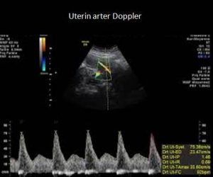
Checking for a low-resistance flow pattern in the umbilical artery, which ensures easy blood flow. Uterine arteries are the blood vessels that carry blood to the uterus. A uterine artery Doppler scan checks whether sufficient blood is reaching the placenta.
The baby needs plenty of nutrients and oxygen to grow at a healthy rate. Therefore, the walls of the uterine arteries need to be flexible enough to allow as much blood to flow through as possible. These small arteries normally enlarge during pregnancy to ensure that more blood can easily reach the uterus, creating a low-resistance flow pattern. Lifestyle factors such as smoking, alcohol consumption, or certain medications can increase the resistance of these arteries and affect blood flow.
To check for Rh sensitization. Blood flow from a blood vessel in the brain can be used to assess the fetus’s health.
Other Information Gained from a Fetal Doppler Scan:
During pregnancy, obstetric Doppler scanning can provide detailed information about blood circulation in the brain, kidneys, heart, umbilical cord, placenta, and uterus. It helps confirm the fetal heart rate. It checks the condition of the umbilical cord, which carries oxygen and nutrients from the mother to the fetus and returns blood lacking nutrients and oxygen.
It monitors the condition of the placenta, which carries all the nutrients needed by the baby, removes waste, supplies oxygen, eliminates carbon dioxide, protects the baby from infections, and secretes the necessary hormones during pregnancy.
With general development screening, the baby’s growth and development can be tracked by measuring length and weight.
What Does Obstetric Doppler Scanning Look For and Why Is It Done?
Uterine artery Doppler scan: Uterine arteries are the blood vessels that carry blood to the uterus. Normally, these arteries are small in size. However, during pregnancy, they increase in size (dilate) to allow more blood to flow through with less resistance. Doppler scanning in this area helps check whether sufficient blood is reaching the uterus.
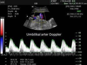
Umbilical artery Doppler scan: In an umbilical artery Doppler scan, blood flow from the baby through the umbilical cord to the placenta is checked. This scan helps ensure that the baby is receiving everything needed from the mother’s placenta. An umbilical artery Doppler scan is performed if the mother is carrying multiple babies or if the baby is growing slower than expected for the gestational age.
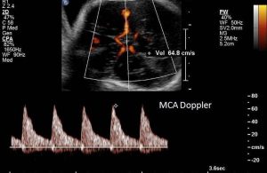
Middle cerebral artery Doppler scan (MCA Doppler scan): If problems are detected during the umbilical artery Doppler scan, an MCA Doppler scan is performed to check blood flow to the baby’s brain.
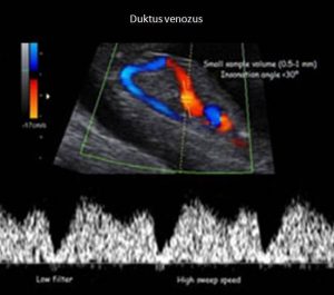
Ductus venosus Doppler scan: This scan evaluates blood flow in the ductus venosus, a blood vessel that carries well-oxygenated blood from the umbilical vein to the baby’s heart. In cases where Doppler abnormalities are present due to fetal growth restriction, blood flow in the ductus venosus will significantly increase.
How Does Doppler Ultrasound Work?
Doppler ultrasound is a painless, non-invasive imaging technique. It uses reflected sound waves to visualize how blood is flowing within the blood vessels. Sound waves reflect off solid objects, including moving blood cells, and measure their speed. The movement of blood cells causes a change in the pitch of the reflected sound waves. This is called the Doppler effect. If there is no blood flow, the pitch does not change.
Beyond the visually striking 3D and 4D images that attract everyone’s attention, the Doppler feature of ultrasound is even more impressive and informative. It is used during pregnancy to study blood circulation in the fetus, uterus, and placenta. Doppler ultrasound uses sound waves to detect the movement of blood, especially red blood cells, in the blood vessels. These sound waves are then reflected back to the ultrasound probe when they contact the red blood cells in the womb. By comparing the characteristics of the sound waves sent by the ultrasound with the characteristics of the reflected sound waves, an ultrasound image is created.
The Doppler principle is similar to the sounds we hear from ambulances or fire trucks in daily life. Imagine an approaching ambulance or fire truck with flashing lights and sirens. Picture how the sound changes as the vehicle gets closer and how the pitch increases, reaching its peak as it passes by, then decreases as the vehicle moves away. In Doppler ultrasound, red blood cells resemble the ambulance vehicle, rushing from one place to another. The “sound” these red blood cells produce depends on their flow speeds. Hence, the term Doppler velocity measurement is used.
During pregnancy, Doppler ultrasound can be used to evaluate maternal blood flow to the uterus or, more commonly, fetal blood flow. Blood flow in the arteries has a different appearance than blood flow in the veins. Venous flow is largely uniform during the cardiac cycle unless the fetus is making breathing movements or there is an adverse clinical condition such as intrauterine growth restriction. However, arterial blood flow is quite different. Regardless of which artery is being evaluated, the speed of the blood moving through the artery will change with each cardiac cycle. The speed will be at its maximum when the heart ejects blood from the left ventricle in a phase called systole. The ventricle then begins to refill in a phase called diastole. During diastole, the blood flow speed gradually decreases, reaching its lowest point at
the end of the filling cycle, called end-diastolic flow, before returning to maximum speed with the next heartbeat. This burst of flow when the heart contracts and pumps out blood is what is felt when someone checks our pulse.
From this, the speed of blood flow moving through the umbilical cord between the baby and placenta can be calculated. Thus, the functionality and health of the placenta and other large blood vessels can be checked. Blood clots, blocked arteries, or decreased blood flow, as well as the risk of preeclampsia, can be identified.
What is the Difference Between a Regular Ultrasound and a Doppler Ultrasound?
A regular ultrasound does not show blood flow velocity, while a Doppler scan shows the blood flow and pressure in the baby’s placenta, umbilical cord, brain, and heart.
Regular ultrasound scans only show the baby’s image, but Doppler allows access to blood flow speed, which helps to determine the amount of oxygen and nutrients the baby is receiving and to hear the heartbeat.
In Doppler ultrasound, the burst of blood flow when the heart contracts and pumps out blood is measured as systolic blood flow (S). Diastole (D), which refers to the heart’s refilling, is measured just before another contraction. The waveform generated by measuring blood flow velocity will have a characteristic sawtooth pattern on the ultrasound screen. The peak of the wave represents systole, and the lowest point represents end-diastolic flow. Depending on clinical conditions, it is possible for there to be no flow in the artery at the end of diastole, known as the absence of end-diastolic flow (AEDF). In AEDF, the waveform will look like sawteeth on the ultrasound screen, but there will be gaps between each tooth.
Blood flow in the umbilical arteries should normally be forward-directed. In extreme clinical conditions such as severe intrauterine growth restriction, arterial blood flow can reverse at the end of diastole. In the case of reversed end-diastolic flow (REDF), instead of “gaps between the teeth,” the waveform will appear as if the “teeth” point both upward and downward. Remember, “teeth” are just a layman’s term for waveforms. Traditionally, downward-pointing waveforms represent blood flow that temporarily moves in the opposite direction of the intended flow. This is called reversed end-diastolic flow (REDF).
A normally functioning placenta should exhibit a low-resistance network of blood vessels through which oxygen, carbon dioxide, electrolytes, nutrients, and waste products are easily exchanged between the mother’s and fetus’s blood vessels. The mother’s blood and the baby’s blood do not mix. However, the exchange of these substances occurs through multiple mechanisms. Increased placental resistance hinders this exchange. Uteroplacental dysfunction is often established in the early weeks of pregnancy, following embryo implantation.
What Are the Clinical Implications of Doppler Findings Such as Intermittent AEDF, Persistent AEDF, and Finally, REDF?
These conditions, if persistent, indicate a worsening intrauterine environment for the developing fetus and present as intrauterine growth restriction. Initially, they appear as increased resistance in Doppler readings, indicating that the intrauterine growth environment is suboptimal. In many patients, the clinical situation may not worsen further and may never progress to reversed end-diastolic flow. These patients typically require biweekly fetal monitoring with Doppler measurements.
However, in other cases, resistance gradually increases to the point of intermittent AEDF or persistent AEDF. Patients who begin to show intermittent AEDF are in a clinical scenario that is often serious enough to require hospitalization. Persistent AEDF or reversed end-diastolic flow indicates that the intrauterine environment has deteriorated to the point where oxygen and carbon dioxide exchange is severely impaired. In cases of reversed end-diastolic flow, oxygen-carbon dioxide exchange is so poor that the fetus may no longer be able to tolerate the environment.
In fetuses with Doppler abnormalities and intrauterine growth restriction, optimal delivery timing depends on the underlying cause and the estimated gestational age. In addition to the umbilical arteries, we can assess the middle cerebral artery in the brain to determine whether the fetus is trying to protect the brain in a low-oxygen environment.