


What is Early Pregnancy Color Doppler Ultrasound?
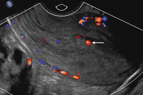
Pelvic color Doppler ultrasonography (CDUS) and testing β-hCG serum levels play a key role in diagnosing early pregnancy. In early pregnancy, color Doppler imaging should primarily be transvaginal. These tests allow differentiation among the diagnostic possibilities of early pregnancy and have contributed to a significant decrease in mortality from ectopic pregnancy.
Normal Development of Early Pregnancy Between 4-8 Weeks of Gestational Age
Gestational age is calculated from the first day of the last menstrual period; however, it is important to know that conception does not occur until about two weeks after ovulation, which is why there is a two-week discrepancy between clinical and histological gestational age. A gestational sac can first be seen on an endovaginal ultrasound at 4.5-5.0 weeks of gestation as a 2-3 mm round intrauterine fluid collection. The mean sac diameter (MSD) growth rate is 1.13 mm per day but is usually variable.
Before confirming that the fluid collection is a true gestational sac by visualizing the yolk sac or embryo, two markers can be used. The intradecidual sign, which is a relatively intact collapsed uterine cavity with an eccentric gestational sac embedded in the echogenic decidua, can be seen as a thin echogenic line and strongly suggests intrauterine pregnancy.
The double sac sign, characterized by the presence of two concentric echogenic rings separated by a thin crescent of endometrial fluid surrounding the fluid collection, is a definitive sign of intrauterine pregnancy. The outer echogenic ring represents the decidua parietalis, and the inner ring represents the decidua capsularis and chorion. The intradecidual sign can appear earlier than the double sac sign because, in the intradecidual sign, the gestational sac is not yet large enough to deform the contour of the uterine cavity, whereas in the double sac sign, the sac protrudes into the endometrial cavity.
The ultrasound appearance of early gestational sacs is variable, and although these two signs strongly suggest early intrauterine pregnancy, they are absent in at least 35% of gestational sacs. Therefore, the absence of these signs does not rule out intrauterine pregnancy. A nonspecific, empty, round intrauterine fluid collection seen in a pregnant patient has a greater than 99.5% chance of representing a gestational sac.
The yolk sac is the earliest gestational sac structure visible on ultrasound. It can typically be seen as an eccentric, round 3-5 mm structure at approximately 5.5 weeks of gestation.
In gestational sacs between 5.0-5.5 weeks, the yolk sac may sometimes appear as two parallel lines representing the front and back walls rather than a separate circle.
The embryo first becomes visible as a 1-2 mm structure adjacent to the yolk sac at approximately 6 weeks of gestation. The embryo is measured from crown to rump, and this measurement, known as the crown-rump length (CRL), is the most accurate method for determining gestational age during the first 12 weeks. The embryo should be visible when the MSD is at least 25 mm.
The embryo is located within the amniotic cavity, and the yolk sac is located within the chorionic cavity. The amniotic membrane is thinner than the yolk sac and can be seen as early as 6.5 weeks of gestation, though it is more easily visualized after 7 weeks.
Between 6.5 and 10 weeks of gestation, there is a linear relationship between the average diameter of the amnion and the CRL, with the amniotic cavity being 10% larger than the CRL. In a normal pregnancy, the chorionic cavity, amniotic cavity, and CRL grow proportionally until around 10 weeks when fetal urine production begins. Fetal urine disproportionately enlarges the amniotic cavity, and by 14-16 weeks, the amnion and chorion fuse, after which the amniotic cavity grows faster than the chorionic cavity.
Cardiac Development in Early Pregnancy Color Doppler Ultrasound
Cardiac pulsation begins in the double endocardial heart tube around the 6th week of pregnancy, allowing cardiac activity to be observed in embryos as small as 1-2 mm. However, the absence of cardiac activity in embryos smaller than 4 mm may still be normal. Cardiac activity should be present in embryos with a CRL of 7 mm or greater, and a failed pregnancy diagnosis can only be confirmed if the embryo is at least 7 mm in length and lacks cardiac activity.
The embryonic heart rate is 100 beats per minute at 6 weeks of gestation and 120 beats per minute at 7 weeks. Embryonic tachycardia, defined as a heart rate of 135 beats per minute or higher before 6 weeks or 155 beats per minute or higher at 7 weeks, is associated with a good prognosis.
Embryonic morphology is nonspecific until about 7-8 weeks when the spine can be visualized. Around the 8th week of pregnancy, the head can be distinguished from the body, and the four limb buds become visible. A notable milestone in development is the rhombencephalon, the developing hindbrain, which becomes visible between 8-10 weeks as an anechoic, round structure within the head. Spontaneous embryonic movement can be seen as early as 8.0-8.5 weeks.
Abnormal Findings in Early Pregnancy Color Doppler Ultrasound
The visualization of early pregnancy signs – the gestational sac at around 5 weeks, the yolk sac at 5.5 weeks, and the embryo at 6 weeks ±0.5 weeks of variation – means that any deviation from this timeline could be indicative of a failed pregnancy. Due to interobserver variability in endovaginal US measurements of CRL, a CRL of 7 mm is required to achieve 100% specificity and positive predictive value for pregnancy failure.
An MSD of 25 mm without an embryo is a valid criterion for pregnancy failure, and an MSD range of 16-24 mm without an embryo is an indicator of suspected pregnancy failure.
Additional findings suspicious for pregnancy failure include an empty amniotic sac, an enlarged yolk sac, and a gestational sac that is small for the size of the embryo. In a normal pregnancy, the amniotic cavity and CRL grow proportionally between 6.5 and 10 weeks. The presence of an “empty amnion” without an adjacent embryo is a sign of poor prognosis and requires Doppler ultrasound follow-up. An irregular gestational sac suggests abnormal intrauterine pregnancy. The presence of a calcified yolk sac indicates that embryonic death likely occurred two or more weeks prior. An enlarged amnion, embryonic bradycardia with a heart rate of 85 beats per minute or less, degenerative hydropic changes in the chorionic villi, and an amorphous embryo between 7-8 weeks are also signs of poor prognosis and require US follow-up.
Early Pregnancy Color Doppler Prices 2026
The prices for early pregnancy color Doppler ultrasound in Istanbul vary from region to region and even from institution to institution within the same region. For information on early pregnancy color Doppler ultrasound prices, you can contact us at 05378576593.