


What Is A Breast Biopsy?
Imaging studies such as mammograms, ultrasounds, and MRIs, often in combination with physical breast examinations, can lead doctors to suspect breast cancer. However, the only way to know for sure is to take a tissue sample from the suspicious area and examine it under a microscope.
A biopsy is a small piece of tissue taken and checked in a laboratory. Breast tissue for a biopsy can be obtained using a special biopsy needle or removed during surgery. It is checked to see if cancer or other abnormal cells are present.
Breast biopsy is used to detect lumps or abnormalities in the breast, often identified through physical examination, mammograms, or other imaging methods. However, these imaging tests don’t always allow determining whether a lump is benign or malignant.
A breast biopsy involves taking a sample of cells from a suspicious area in the breast identified through imaging methods and examining them under a microscope for diagnosis. This biopsy is less invasive than surgical tissue sampling. A breast biopsy is not done to remove the entire lump.
An ultrasound-guided breast biopsy involves taking a tissue sample from a lump seen on the ultrasound, guided by the ultrasound imaging.
A pathologist (a doctor specialized in diagnosing disease) examines the tissue sample to see if cancer cells are present. If cancer is found, the pathologist can evaluate its characteristics. The biopsy results in a report detailing all of the pathologist’s findings.
Why Is A Breast Biopsy Done?
To check a lump or swelling in the breast that can be felt (palpable),
To evaluate a problem seen on a mammogram, such as small calcium deposits (microcalcifications) or a fluid-filled lump (cyst),
To assess nipple problems such as bloody discharge,
To determine whether a breast lump is cancerous (malignant) or benign (non-cancerous).
A lump or area of concern in the breast may be caused by cancer or a less serious condition.
A breast ultrasound may indicate abnormalities such as:
A suspicious lump, structural distortion in breast tissue, or abnormal areas of tissue change, warranting an ultrasound-guided breast biopsy.
Before A Breast Biopsy, Inform Your Doctor If:
If you have any allergies,
If you have used aspirin in the past seven days,
If you are taking blood-thinning medications (anticoagulants).
How Is A Breast Biopsy Performed?
There are several types of breast biopsy procedures. The type you undergo will depend on the location and size of the breast lump or area of concern.
Different techniques may be used for the biopsy, and your doctor will likely try to use the least invasive procedure possible—one involving the smallest incision and minimal scarring. However, the choice of procedure depends on your personal situation. A biopsy can be performed by inserting a needle through the skin into the breast to extract a tissue sample.
Biopsies may be performed under local or general anesthesia. Local anesthesia involves injecting medication to numb the breast. You will be awake but won’t feel any pain.
Medical guidelines suggest that about 90% of biopsies should be needle biopsies, the least invasive procedure. However, studies show that about 70% of breast biopsies are surgical. This means that many women undergo unnecessary surgeries, and those diagnosed with cancer need a second surgery to remove the cancer.
Types of breast biopsies include:
Ultrasound-guided breast biopsy types
Fine Needle Aspiration Biopsy
Fine needle aspiration is the simplest form of breast biopsy and can be used to assess a lump detected during a clinical breast exam. For the procedure, the patient lies on their back. While visualizing the lump with an ultrasound probe in one hand, the doctor guides a very thin needle into the lump with the other hand.
The needle is attached to a syringe that collects a cell sample. Fine needle aspiration is a quick way to differentiate between a fluid-filled cyst and a lump, potentially avoiding more invasive biopsy procedures.
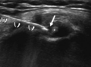
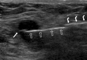
Tru-cut (Core) Biopsy:
This type of breast biopsy is used to evaluate a breast lump detected on a mammogram or ultrasound or during a breast exam. The radiologist often uses ultrasound guidance to extract tissue samples from the lump using a tru-cut needle. Several samples, each the size of a grain of rice, are collected and analyzed pathologically to determine tissue characteristics.
Ultrasound-guided tru-cut (core) biopsy: The biopsy needle is positioned using ultrasound, a method that produces detailed images of the body’s structures using high-frequency sound waves. You will lie on your back during the procedure, and the radiologist will locate the lump in your breast using the ultrasound probe, positioning the needle for the biopsy.
Both fine needle aspiration and core needle biopsy offer rapid results without significant discomfort or scarring, providing an opportunity to discuss treatment options with your doctor before undergoing any surgery.
After a breast biopsy, you will leave with only bandages over the biopsy area. You can rest for the remainder of the day or return to your normal activities. After a breast biopsy, you can take pain relievers to manage any discomfort and apply cold compresses to the biopsy site to reduce swelling.
Vacuum-Assisted Breast Biopsy
Vacuum-assisted breast biopsy is a newer method for performing a breast biopsy. Unlike the tru-cut biopsy, which involves inserting a needle through the skin several times, vacuum-assisted biopsy can remove more tissue.
For a vacuum-assisted breast biopsy, you will lie face down on a special examination table with round openings for your breasts. A local anesthetic injection is administered to numb the breast. Guided by mammography (stereotactic biopsy) or ultrasound, the radiologist inserts the probe into the suspicious area of the breast. A vacuum then pulls tissue into the probe, where a rotating cutting device removes a tissue sample and transfers it to a collection area. The radiologist can rotate the probe to take additional samples from the suspicious area, repeating this process as needed to sample all relevant areas thoroughly.
In some cases, a small metal clip is placed in the biopsy area to mark the location for future biopsies. This clip remains in the breast and does not cause pain or harm.
Incisional Breast Biopsy
An incisional biopsy is more like traditional surgery. After administering an anesthetic injection, the surgeon uses a scalpel to remove a piece of tissue and cut through the skin.
As with needle biopsies, if the surgeon cannot feel the suspicious area, mammography or ultrasound may be needed to guide the surgeon to the correct spot. Your doctor may also use a procedure called needle localization. Under mammographic or ultrasound guidance, the radiologist inserts a small hollow needle into the abnormal area through the breast skin. A small wire is inserted through the needle into the affected area. The needle is then removed, and the doctor uses the wire as a guide to locate the correct spot for the biopsy.
If a needle biopsy is inconclusive (i.e., the results are unclear or uncertain), or if the suspicious area is too large to be easily sampled with a needle, your doctor may recommend an incisional biopsy. Since it is a surgical procedure, an incisional biopsy is more invasive, leaves a scar, and may take longer to heal than a needle biopsy.
Excisional Breast Biopsy
An excisional biopsy is surgery to remove the entire suspicious area of tissue from the breast. In addition to removing the suspected cancer, the surgeon usually also removes a small margin of normal tissue surrounding it,