


Parotid Gland Ultrasound is considered the first imaging method for evaluating lymph nodes and soft tissue diseases in the head and neck, including the major salivary glands. The results of ultrasound examination alone can suggest a definitive diagnosis or provide important differential diagnostic data.
As the head and neck region has a complex anatomical structure, solid knowledge of sonographic anatomy and spatial relationships is crucial for reliably performing the examination. In addition, understanding the sonographic features of the most common diseases in this area is also essential.
Normal Anatomy
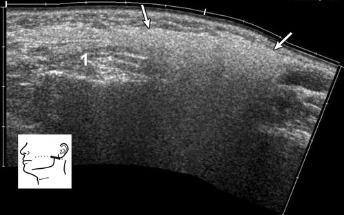
The parotid gland is located in the retromandibular fossa, in front of the ear and the sternocleidomastoid muscle. The boundary between the superficial and deep parotid lobes is formed by a plane in which the facial nerve and its branches are located. The branches of the facial nerve are not visible on ultrasound. The deep parotid lobe can only be partially visualized on ultrasound.
The normal echogenicity of all major salivary glands, including the parotid gland, is generally homogeneous and varies between highly echogenic and mildly echogenic compared to adjacent muscles.
The echogenicity of the parotid gland depends on the amount of intraglandular fat tissue. Salivary glands with high fat content are more echogenic than surrounding muscles. The length of Stenson’s duct usually varies between 3 and 5 cm. During ultrasound examination, the non-dilated duct is generally invisible.
Lymph nodes may be found within the parenchyma of the parotid gland. They are primarily localized in the upper and lower poles of the gland. The presence of a hyperechoic hilum is an important criterion for the normalcy of parotid lymph nodes.
Acute Inflammation
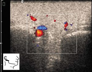
Acute inflammation typically causes painful swelling of the salivary glands, usually bilaterally. In acute inflammation, the salivary glands enlarge and become hypoechoic. They may be heterogeneous, containing numerous small, oval, hypoechoic areas. Enlarged lymph nodes with increased central blood flow can be seen in acute inflammation of the salivary glands.
Abscess
Abscess formation may occur during acute sialadenitis. On ultrasound, abscesses appear as hypoechoic or anechoic lesions with ill-defined margins and posterior acoustic enhancement.
Chronic Sialadenitis
Chronic sialadenitis is clinically characterized by intermittent swelling of the gland, which may or may not be associated with food. In chronic inflammation, the salivary glands are of normal size or smaller, hypoechoic, and heterogeneous. Typically, there is no increase in blood flow on ultrasound.
Neoplasms
Salivary gland neoplasms are relatively rare. Most are benign (70%-80%) and are found in the parotid glands (80%-90%).
Benign Neoplasms
The most common benign neoplasms of the major salivary glands are pleomorphic adenomas and Warthin’s tumors. They clinically present as slow-growing, painless masses.
However, small lesions may be incidentally detected on ultrasound. When their ultrasound appearances are analyzed, many common features can be found, but a definitive differential diagnosis between benign and malignant tumors is often not possible with US alone.
Pleomorphic adenoma
Malignant Neoplasms
The most common malignant neoplasms of the salivary glands are mucoepidermoid carcinoma and adenoid cystic carcinoma. Squamous cell carcinoma, acinic cell carcinoma, and adenocarcinoma are less common.
Unlike benign salivary neoplasms, malignant tumors can be detected as rapidly developing, tender or painful masses upon palpation, and may cause paralysis or paresis of the facial nerve.
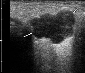
The typical US features of poorly differentiated or underdeveloped malignant neoplasms of the salivary glands are similar to those in other organs or tissues. The ultrasound characteristics of malignant neoplasms include irregular shape, ill-defined, blurred edges, and a hypoechoic, heterogeneous structure.
However, malignant tumors can also be homogeneous and well-circumscribed. The internal structure of a malignant tumor on ultrasound may not only be solid but also cystic, or the cyst may have a nodule within its wall. Malignant tumors can have a lobulated shape, similar to pleomorphic adenomas.
Parotid Gland Ultrasound Prices 2026
Parotid gland ultrasound prices 2026 – you can contact us immediately to get information.
How Much Does It Cost?
Dr. Abdullah Cevahir
Radiology Specialist
Hekimoğlu Imaging and Diagnostic Center