


Neck Ultrasound is the first imaging method following a clinical examination in the neck region.
It provides valuable diagnostic information with a high degree of diagnostic accuracy.
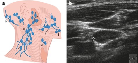
Lymph Node Ultrasound Findings
They appear sharply defined and oval in configuration with a homogeneous hypoechoic appearance. They have a central echogenic hilum structure. Typically, the short axis is less than 1 cm. The ratio of the short axis to the long axis is less than 0.7.
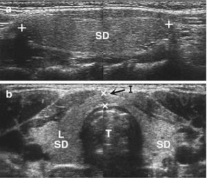
Thyroid Gland
Tubular anechoic structures representing vessels are seen within the parenchyma, which is slightly hypoechoic and homogeneous compared to surrounding tissues. The echogenicity of the parenchyma increases slightly with age.
The anterolateral border is convex in shape (a concave shape indicates an increase in size and can appear locally only in the case of regional infiltration).
In older children, slight cystic changes and nodular-patchy parenchymal irregularities may be normal.
The size for each lobe is calculated using the ellipsoid formula (Length × Width × Thickness × 0.48), and the total volume = the sum of the left and right lobes.
Thyroid volume changes with age and shows geographic regional variation.
Thyroid parenchymal lesions: A simple cyst appears as an anechoic, thin-walled structure on ultrasound. A complex cyst is septated with thick walls and internal echogenicities. Solid nodules with benign features, such as adenomas, tend to be echogenic with a homogeneous internal structure. The main common points of nodules with malignant features are their rapid growth, more hypoechoic and heterogeneous internal structure, irregular indistinct borders, and sometimes containing microcalcifications.
Thyroglossal Duct Cysts
This is a duct extending between the root of the tongue and the thyroid gland, located near the midline of the neck. In most cases, there is a connection between the cyst and the hyoid bone. On ultrasound, a thyroglossal duct cyst appears
as a typical anechoic cystic lesion. Sometimes they are multicystic and may contain components of echogenic thyroid tissue. In case of infection, the cyst becomes painful, and the fluid inside the cyst exhibits a heterogeneous echo structure due to pus. In 1% of thyroglossal duct cysts, carcinoma can develop, typically papillary adenocarcinoma. In such cases, the cyst grows, and solid tumor tissue fills the cystic lumen.
Parotid, Submandibular, and Sublingual Glands
Parotid gland: It has a slightly irregular, somewhat heterogeneous, lobular, and hilar-vascularized echogenic parenchyma. The parotid duct is only visible when obstructed and dilated.
Submandibular / sublingual gland: Similar echogenicity to the parotid, but without lobules, potentially less irregular. Hilar structures are less prominent compared to the parotid.
Branchial Cleft Cysts
These are most commonly located in the ventrolateral region of the carotid bifurcation. They develop from the first or second branchial clefts and have variable echogenicity. They may be anechoic,
but most cysts are moderately echogenic and homogeneous. There is a granular echo pattern caused by cellular debris or cholesterol crystals within the cyst. When inflammation is present, the fluid content becomes heterogeneous and echogenic.
Neck Ultrasound Prices 2026
Neck ultrasound prices 2026 – you can contact us immediately to get information.
How Much Does It Cost?
Dr. Abdullah Cevahir
Radiology Specialist
Hekimoğlu Imaging and Diagnostic Center