


What is Breast Ultrasound?
Breast ultrasound is a commonly used imaging technique for screening breast masses and other breast abnormalities. Ultrasound uses high-frequency sound waves to create detailed images of the breast. Unlike X-rays and CT scans, ultrasound does not use radiation and is considered safe for pregnant women and breastfeeding mothers.
Why is Breast Ultrasound Performed?
The primary use of breast ultrasound is to help diagnose breast abnormalities detected during physical examination by a doctor and to characterize potential abnormalities seen in mammography or breast magnetic resonance imaging (MRI).
Ultrasound imaging can help determine whether the abnormality is solid (which could be non-cancerous tissue or a cancerous tumor), fluid-filled (like a benign cyst), or both cystic and solid.
Doppler ultrasound is used to evaluate blood flow in breast lesions.
Mammography is the only screening tool for breast cancer known to reduce deaths due to breast cancer through early detection. However, mammograms do not detect all breast cancers.
Some breast lesions and abnormalities may not be visible on mammography or may be difficult to interpret. Dense breasts have a lot of glandular and connective tissue with little fatty tissue, making cancer detection more challenging.
Many studies have shown that ultrasound and magnetic resonance imaging (MRI) can help support mammography by detecting breast cancers that are not visible on mammograms. Your doctor will tell you which of these tests is appropriate for you. MRI is more sensitive than ultrasound in depicting breast cancer.
If MRI is performed, ultrasound may be used afterward to characterize abnormalities seen on the MRI and to guide biopsies of these abnormalities. When ultrasound is used for screening, abnormalities not visible on mammography can be identified. Most abnormalities detected by screening ultrasound are not cancer.
Ultrasound may be used for breast cancer screening in:
Individuals at high risk for breast cancer who cannot undergo breast MRI.
Pregnant women or those who cannot be exposed to X-rays.
Increased breast density – when breasts have a higher proportion of dense glandular and connective tissue compared to fatty tissue.
How Do I Prepare for a Breast Ultrasound?
Breast ultrasound does not require any special preparation.
It is important to avoid applying powder, lotion, or other cosmetic products to your breasts before the ultrasound, as this may affect the accuracy of the test.
Breast Ultrasound Results
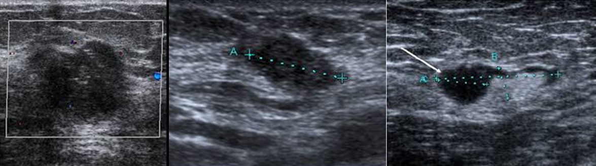
The images produced by a breast ultrasound are black and white. Cysts, tumors, and masses will appear as darker areas on the image.
A dark spot on your ultrasound does not necessarily mean you have breast cancer. In fact, most breast cysts are benign. Several conditions can cause benign cysts in the breast, including:
- Adenofibroma, a benign tumor of breast tissue.
- Fibrocystic breasts, which are painful and cystic due to hormonal changes.
- An intraductal papilloma, a small, benign tumor of the milk duct.
- Fat necrosis of the breast, bruised, dead fat tissue that causes swelling.
If your doctor detects a mass that requires further testing, an MRI may be performed first, followed by a biopsy. Biopsy results will help determine whether the cyst is malignant or benign.
Ultrasound-Guided Breast Biopsy
If a suspicious mass is detected in your breast, your doctor may recommend a breast ultrasound. Ultrasound can help determine whether the mass is a fluid-filled cyst or a solid tumor. It also helps determine the location and size of the mass.
While breast ultrasound can be used to evaluate a mass in your breast, a tissue sample (biopsy) is required to definitively determine whether the mass is cancerous.
In addition to identifying breast abnormalities, breast ultrasound can be used for women who need to avoid radiation exposure, such as:
Women under 25 years of age, pregnant women, breastfeeding women, women with silicone breast implants.
Breast Ultrasound Prices 2026
For information about breast ultrasound prices in 2026, you can contact us immediately.
Dr. Abdullah Cevahir
Radiology Specialist
Hekimoğlu Imaging and Diagnostic Center