


Upper Extremity Arterial Doppler Ultrasound
The arterial system of the upper extremity requires a different diagnostic approach compared to that used for the lower extremity. While the progression of focal atherosclerosis or acute arterial embolism is almost always the cause of symptomatic disease in the lower extremity, upper extremity arterial disease is more complex. Pathologies to consider when evaluating upper extremity arteries include mechanical compression at the thoracic outlet, vasospasm in the digital arteries, thrombus in the hand or wrist due to trauma, arteritis, aneurysms, or emboli from the heart or proximal arm.
Anatomy of the Upper Extremity Arterial Doppler
The upper extremity arterial system originates from the aortic arch. On the right, there is a common trunk called the innominate or right brachiocephalic artery, which then divides into the right common carotid artery (CCA) and subclavian artery. On the left, the subclavian artery arises directly from the aortic arch. The subclavian artery continues to the lateral edge of the first rib, where it becomes the axillary artery. The axillary artery passes under the pectoralis minor muscle, crosses the teres major muscle, and then becomes the brachial artery. The brachial artery continues down the arm to divide into the radial, ulnar, and interosseous (or median) arteries just below the elbow. The radial and ulnar arteries are the dominant branches continuing to the wrist.
The radial and ulnar arteries typically unite in the hand through the superficial and deep palmar arches, which then supply the fingers via common palmar digital arteries and communicating metacarpal arteries.
How is the Upper Extremity Arterial Color Doppler Exam Performed?
The upper extremity arterial exam normally begins with the proximal subclavian artery. The entire course of each major artery, including the subclavian, axillary, brachial, radial, and ulnar arteries, is imaged.
Color Doppler ultrasound is used to detect blood flow in the vessels and provide the examiner with information about the speed and direction of blood flow. Normal upper extremity Doppler waveforms are triphasic; however, waveforms may change in response to environmental factors, such as temperature or maneuvers like clenching the fist, especially when measured near the hand.
Color Doppler Evaluation of Upper Extremity Arterial Pathologies
Clinically significant atherosclerotic plaque usually develops in the proximal subclavian arteries and sometimes in the axillary arteries. The primary effect is a reduction in blood flow due to stenosis or occlusion, which can lead to arm ischemia. Atherosclerotic obstruction in more distal arteries, such as the brachial, radial, and ulnar arteries, is less common; however, distal arteries may become blocked secondary to low-flow conditions or embolization.
While stenosis of the proximal upper extremity arteries is most commonly caused by atherosclerosis, other pathologies include vasculitis, trauma, or thoracic outlet compression. Stenosis that narrows the lumen diameter by 50% or more is considered hemodynamically significant, as it reduces blood flow. Such stenosis is identified by an increase in peak systolic velocities, and in the case of severe stenosis or occlusion, a dampened (tardus-parvus) waveform distal to the high-grade stenosis or occlusion may be observed. Additionally, high-grade arterial stenosis or occlusion causes a general reduction in blood flow velocities distal to the obstruction. Stenosis is considered hemodynamically significant when it causes a doubling of peak systolic velocities (compared to pre-stenotic velocities), indicating ≥50% lumen narrowing. More severe stenosis will further increase both systolic and diastolic velocities.
Color Doppler imaging of stenosis can identify (1) narrowing of the arterial lumen; (2) changes in color flow signals consistent with increased blood flow velocities at the stenosis; and (3) a post-stenotic turbulent flow pattern. With arterial occlusion, color and power Doppler may show a high-resistance flow pattern with no detectable flow in the occluded vessel lumen or Doppler spectral waveforms, often accompanied by decreased peak systolic velocities. Distal to the obstruction, a dampened, monophasic flow pattern is seen.
Arterial thrombosis can occur distal to a critical stenosis or result from embolization, trauma, or thoracic outlet compression. The result can be either complete or partial occlusion. Thrombus or vasculitis may be visualized directly with gray-scale imaging, but color Doppler imaging is used to determine vessel patency and assess the degree of vessel recanalization following thrombolysis.
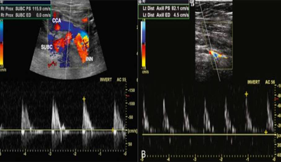
If there is a significant difference in arm blood pressures (e.g., 20 mm Hg), color Doppler evaluation should be extended to check for subclavian steal. Subclavian steal syndrome is a condition in which there is severe stenosis or occlusion in the subclavian artery proximal to the origin of the vertebral artery. The pressure drop caused by the occlusion results in the subclavian artery being supplied by the ipsilateral vertebral artery. Subclavian steal can cause symptoms by reducing blood flow to the affected arm.
Thoracic Outlet Syndrome
Thoracic outlet syndrome is a potential cause of upper extremity circulatory problems, such as a cold, painful, or numb extremity that varies with limb position. It is important to diagnose or rule out thoracic outlet vascular compression early. The thoracic outlet is bordered by the clavicle, first rib, and scalene muscles. Thoracic outlet syndrome is a common problem caused by compression of the subclavian vessels and/or the brachial plexus as they exit the chest. Compression at the thoracic outlet can partially or completely obstruct blood flow when the arm is in certain positions. Over time, repeated vascular irritation can damage the artery or vein, leading to intimal injury or thrombus formation. Arterial emboli from thrombi in this region can travel distally to other parts of the upper extremity. Therefore, whenever emboli are found, they should be considered to have potentially originated from the subclavian or axillary arteries.
Most patients with uncomplicated thoracic outlet syndrome (without thrombus, plaque, or aneurysm) do not experience arm symptoms unless the arm is in a specific position. The discomfort the patient feels is likely due to nerve compression rather than temporary loss of blood flow caused by arterial compression, at least in the early stages. The patient describes pain, numbness, or loss of sensation when the upper extremity is in a predictable and reproducible position, such as brushing hair or driving with hands on the steering wheel. Symptoms disappear immediately after the arm is repositioned.
Upper Extremity Arterial Color Doppler Prices 2026
Upper extremity arterial color Doppler prices may vary depending on the region, especially in Istanbul. For detailed information about upper extremity arterial color Doppler prices, please call us at 02126321060 or 05359214295.