


Superficial tissue ultrasound refers to the examination of palpable lumps or swellings in any part of the body using an ultrasound device.
Ultrasound is the primary screening tool in investigating superficial tissue lesions. Compared to other imaging methods, ultrasound is a safe (non-ionizing), portable, easily repeatable, and inexpensive imaging technique. Ultrasound is an excellent method for determining the nature (cystic or solid) of a mass lesion and its anatomical relationship with adjacent structures.
Non-tumoral superficial lesions
The most common non-tumoral lesions are benign cysts, trauma-related, reactive, and inflammatory lesions. These lesions include synovial cysts, ganglion cysts, tenosynovitis, epidermoid cysts, and masses showing cystic transformation (abscess, necrosis, hematoma).
Benign cysts
Benign cystic masses often appear as localized swellings near or adjacent to joints. Synovial cysts are often associated with joints. The most common synovial cysts are popliteal or Baker’s cysts.
Baker’s cyst
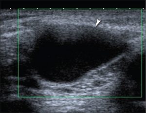
Ganglion cysts can occur anywhere but are most often seen in the hand, wrist, foot, and ankle. The most common cause of a palpable lump in the hand and wrist is a ganglion cyst. They are often seen in young women, and about 10% of patients have a history of trauma. Ganglions can extend into the joint space through a narrow neck. Ganglions are typically anechoic on ultrasound. They do not show vascularization on color Doppler ultrasound.
Ganglion cyst
Epidermoid cysts are clinically most often seen as epithelial-lined cysts that appear in hair-bearing areas of the human body. On sonography, epidermoid cysts are typically hypoechoic. A lamellar sonographic pattern is diagnostic in epidermoid cysts.
Post-traumatic masses
Muscle tears, hematomas, and tendon tears are typically identified through clinical history and physical examination. Partial muscle tears can be displayed as distinct masses, and secondary muscle hypertrophy can be seen in chronic cases. Tendon ruptures may occur spontaneously or due to trauma in patients with rheumatoid arthritis (RA) or other rheumatological conditions with chronic synovitis.
Myositis ossificans may occur secondary to trauma or surgery. Calcifications in myositis ossificans appear hyperechoic and are located peripherally on ultrasound images.
Reactive and inflammatory masses
Inflammatory masses may have infectious or non-infectious origins. Non-infectious reactive and inflammatory masses include neuromas, giant cell tumors of the tendon sheaths, foreign body granulomas, and rheumatoid nodules. Infectious inflammatory masses include abscesses, pyomyositis, or cellulitis.
Neuromas are proliferative responses to nerve damage. On ultrasound, neuromas appear as hypoechoic nodules and typically show minimal vascularity on color Doppler ultrasound.
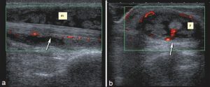
Giant cell tumors of the tendon sheath are the second most common mass in the hand after ganglion cysts. They tend to appear on the volar side of the fingers. On ultrasound, they appear as hypoechoic masses adjacent to joints and are highly vascular structures on Doppler ultrasound.
Foreign body granulomas present as hard and painful swellings. Ultrasound is superior to conventional radiography in detecting radiolucent foreign bodies (such as wood fragments). Foreign bodies usually appear hyperechoic and are surrounded by a hypoechoic halo. Color Doppler shows increased vascularization around the foreign body.
Rheumatoid nodules are found in approximately 20% of patients and appear as elongated hypoechoic nodules adjacent to or within tendons.
Abscesses and cellulitis present with similar clinical findings. Differential diagnosis can be easily made with Doppler ultrasound. Abscesses usually appear as complex cystic lesions with irregular walls, septations, debris, or internal echogenicity.
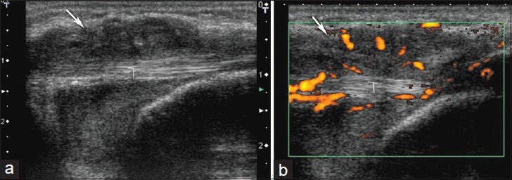
Tenosynovitis is inflammation of the tendon sheath due to various etiologies. Rheumatoid arthritis is one of the most common inflammatory diseases that cause tenosynovitis. Acute suppurative tenosynovitis usually occurs in cases of penetrating injury. Tenosynovitis can be diagnosed sonographically by fluid distending the tendon sheath and/or thickening of the sheath.
Scar (abdominal wall) endometriosis
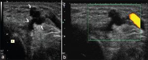
In patients who have undergone a cesarean section or pelvic surgery, scar (abdominal wall) endometriosis can be easily diagnosed based on history. The mass becomes more prominent in the area of the surgery during the menstrual cycle. On ultrasound, endometrial implants usually appear hypoechoic but can also appear hypervascular or avascular on Doppler ultrasound.
Superficial neoplastic masses
Superficial soft tissue tumors can be evaluated with ultrasound and Doppler ultrasound in terms of echogenicity, size, vascularity, quantity, and their relationship with adjacent structures.
Benign neoplastic masses
Lipomas
Lipomas are benign soft tissue masses commonly located subcutaneously, intramuscularly, or intermuscularly. On ultrasound, they appear hypovascular, homogeneous, and predominantly hyper-isoechoic to surrounding fat. They can appear as encapsulated masses within subcutaneous fat tissue.
Fibromatosis
Superficial fibromatosis is a benign condition characterized by fibrous proliferations, typically presenting as slow-growing subcutaneous lesions. On ultrasound, they appear hypoechoic and show variable vascularity.
Extra-abdominal desmoid tumors
Extra-abdominal desmoid tumors are locally aggressive fibroblastic tumors arising from connective tissue of the muscle or fascia of extra-abdominal muscles. On ultrasound, they appear as infiltrative, firm, and heterogeneous masses.
Myxomas
Soft tissue myxomas may be associated with fibrous dysplasia. On ultrasound, they appear as soft, fluctuant hypoechoic masses. They are encapsulated and often contain cystic components.
Nerve sheath tumors
Benign nerve sheath tumors include schwannomas and neurofibromas. Schwannomas tend to appear as hypoechoic, round, and well-defined masses with an eccentric cystic component, narrowing distally on ultrasound.
Vascular malformations
Hemangiomas are common vascular malformations and account for 7% of all soft tissue masses. They are more common in women. Hemangiomas are the most commonly diagnosed soft tissue tumors in the pediatric age group. Ultrasound may reveal prominent vascular channels, phleboliths, and fat.
Malignant masses
Malignant superficial soft tissue masses can be primary or metastatic neoplasms, and the incidence of sarcomas increases with age. On ultrasound, malignant soft tissue tumors typically appear hypoechoic and hypervascular and are well-defined. Cystic components, necrotic areas, and dystrophic calcifications may also be seen.
Superficial Tissue Ultrasound Prices 2026
For information on superficial tissue ultrasound prices for 2026, you can contact us immediately.
What is the cost?
Dr. Abdullah Cevahir
Radiology Specialist
Hekimoğlu Imaging and Diagnostic Center