


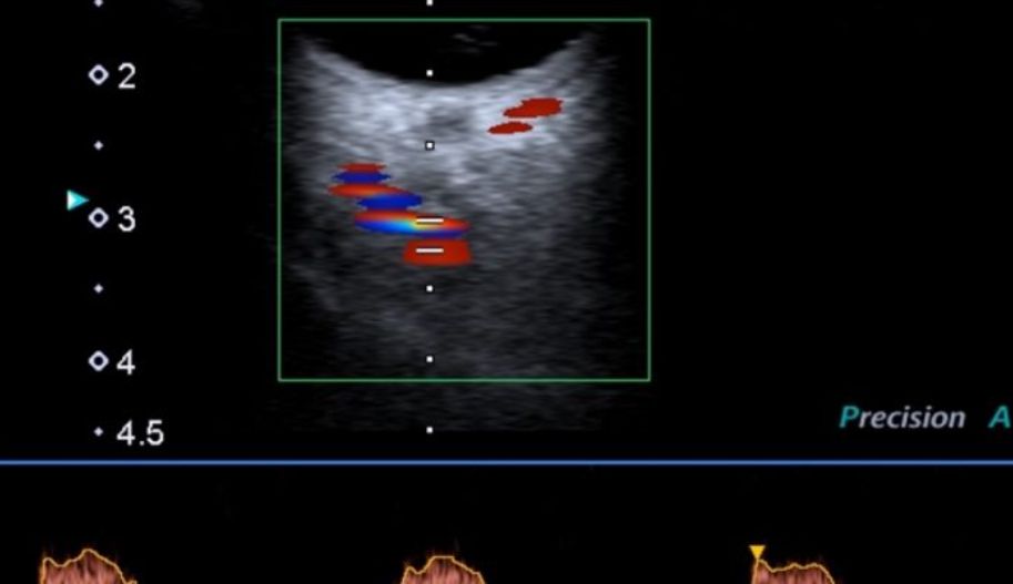
Lower Extremity Arterial Doppler Examination
What is Leg (Lower Extremity) Arterial Doppler Ultrasound?
The arterial Doppler examination of the legs is an imaging method used to evaluate blood circulation and check for blockages or narrowing in the leg arteries.
Peripheral Circulation
The function of blood circulation is to transport nutrients and oxygen to the body’s cells and organs. When the heart pumps blood, the arteries act as highways to carry the blood away from the heart. To keep body cells alive and healthy, the arteries must remain open. “Peripheral” refers to the arteries that run along the arms and legs.
Atherosclerosis
The inner wall of a healthy artery is smooth and strong, allowing blood to flow freely. As we age, the arteries can be affected by atherosclerosis or “hardening of the arteries.” The inner surface of the arteries becomes thickened and rough due to cholesterol or fat buildup, similar to rust forming in a pipe. This buildup is called “plaque,” and it can cause the artery to narrow or even close entirely, reducing or stopping blood flow. Atherosclerosis can occur in any artery, but it most often affects those in the heart, neck, and legs.
Symptoms
Symptoms depend on the amount of collateral arteries that develop to take over the work of the diseased arteries. These collateral arteries are like side roads or detours people use to avoid traffic jams on the main highway.
Claudication Pain – Blockages in the arteries of the lower extremities can cause intense pain, especially during walking. The pain typically subsides with rest. Claudication, commonly referred to as walking pain, can lead to movement limitations. The pain is usually felt in the calf, hip, foot, and thigh areas. It worsens while climbing stairs or slopes and is a key symptom that starts with walking and stops with rest.
The location of the blockage affects where the pain is felt. Blockages in arteries above the groin area may cause pain in the thigh and calf, while blockages in the arteries near the knee may result in foot pain. If there is a blockage in the aorta or iliac arteries, the pain may be felt in the hip area.
Rest Pain – This usually indicates advanced arterial disease. Severe or persistent pain or a burning sensation in the toes, heel, or foot can be very painful. Keeping the leg and foot down for a short time may provide temporary relief (gravity helps blood flow).
Tissue Necrosis (Gangrene) – This is the most serious stage of arterial disease. As the disease worsens, wounds may not heal, and the skin can easily become injured and begin to break down, leading to gangrene.
Risk Factors
Heart disease, high blood pressure, smoking, diabetes, high cholesterol, a family history of vascular disease, lack of exercise
Diagnosis
If your doctor suspects peripheral arterial occlusive disease, they may request an arterial color Doppler examination to confirm the diagnosis.
Anatomy and Pathophysiology of Peripheral Arterial Disease
Peripheral arterial disease is grouped based on the location of the lesion: aortoiliac, femoropopliteal, and crural (tibiopedal). Aortoiliac disease involves the infrarenal abdominal aorta, common iliac arteries, internal iliac arteries, and external iliac arteries, found proximal to the inguinal ligament or at the deep circumflex iliac artery.
Femoropopliteal disease involves the main femoral arteries, deep femoral arteries, superficial femoral arteries, and popliteal arteries. Crural disease involves the anterior tibial, posterior tibial, peroneal, dorsalis pedis, and plantar arteries.
Arterial Doppler Waveform
A continuous wave Doppler velocity detector detects the Doppler shift from sound waves reflecting off moving red blood cells. The B-mode component of duplex ultrasonography ensures correct angle placement between 30° and 70°. A normal lower extremity arterial Doppler flow pattern is triphasic, consisting of a sharp upstroke and peak systolic component, an early diastolic component where flow reverses, and a late diastolic forward flow component.
Distribution of Peripheral Arterial Disease
Peripheral arterial disease can affect an isolated segment of the aortoiliac, femoropopliteal, or crural vascular system, or it may have multisegmental involvement. Crural segments are more commonly affected in diabetic patients, while aortoiliac disease is more frequent in those with a history of smoking.
Aortoiliac Disease
Aortoiliac disease typically refers to atherosclerotic disease affecting the infrarenal abdominal aorta, common iliac, internal, and external iliac arteries.
Femoropopliteal Disease
Femoropopliteal disease affects the main femoral artery, deep femoral artery, superficial femoral artery, and popliteal artery. It typically presents as claudication at the level of the thigh or calf.
Crural Disease
Crural disease involves the anterior tibial, posterior tibial, peroneal, dorsalis pedis, and plantar arteries. Claudication in the foot is rare but typically associated with tibial and peroneal artery involvement.
Lower Extremity Arterial Color Doppler Ultrasound Prices 2026
For information on **Lower extremity arterial color Doppler ultrasound prices 2026**, you can contact us at 05359214295.