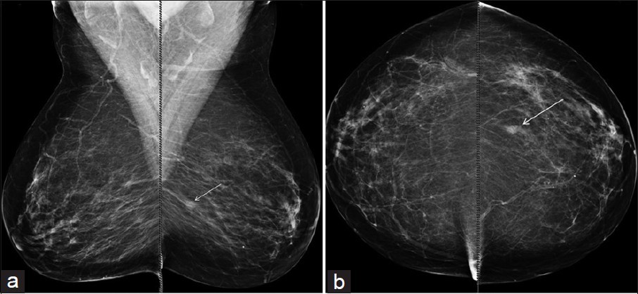
What is a mammogram?
Mammography, a technique that uses low doses of X-rays to image the breast, is an important tool for detecting breast diseases. During a mammogram, your breast is compressed between two flat panels to get a more detailed image. Dec. The radiology technician positions the breast to get the best possible images. Our radiology technicians will help our patients to avoid discomfort as much as possible during the procedure. For the most part, images of each breast are taken in two plans. These images are then interpreted by the radiologist.
Screening mammography is an imaging examination performed once a year in women who do not have significant breast problems. if you are 40 years or older and do not have breast problems, we strongly recommend that you have a mammogram once a year. A mammogram can show changes in the breast long before these changes are felt by a woman or her doctor. screening mammograms performed once a year from the age of 40 have been linked to a dramatic increase in breast cancer survival rates.
A mammogram performed to assess a breast problem, such as a mass in the chest, thickening, swelling, pain, nipple discharge, or changes in the skin, is a diagnostic mammogram. In this type of mammogram, we can get additional close-up images that focus on the abnormal area. Oct.
Digital Mammography is a very valuable imaging method widely used in radiology. A mammogram is actually a kind of X-ray that uses X-rays.
Very detailed, high-resolution images are obtained thanks to filtered X-rays with slightly different energy compared to normal X-rays and a small focus size (the adjustable aperture through the X-ray tube is called focus, small focus diameters such as 0.1-0.3 mm are used in mammography ).
Mammography Saves Lives
With a rate of 1 out of every 8 women, breast cancer is the most common cancer in women. Every year, 40 thousand women lose their lives from breast cancer. 75 Percent of women who get breast cancer are at “average risk,” that is, people who do not have a family history of the disease or other high-risk factors. 1 out of every 6 breast cancers occurs in women aged 40-49 Dec.
The introduction of annual mammography screenings for over-40s has helped reduce breast cancer. Losses due to breast cancer have decreased by more than 40 percent. A mammogram can even detect too many tumors to be felt. Small tumors can be treated more easily and more effectively.
How Do I Prepare for a Mammogram?
If you usually complain of breast pain or tenderness in the days before menstruation, you may want to consider scheduling your mammogram appointment for a time other than this premenstrual time. The best time for mammography is the first 5 days after the end of menstruation.
Do not apply deodorant, body powder or lotion to your breast or armpit areas on the day you are going to have a mammogram. These can cause artifacts in mammography images.
Since you will need to take off your clothes from the waist up, it will be appropriate to choose comfortable and two-piece clothes that may be suitable for this situation.
If you have reports and CDs of mammograms, breast ultrasounds, or breast MRI images that you have had done at other centers, bring them. These will be uploaded to our system so that the radiologist can compare them. This is extremely important, because a change from previous images can be a sign of breast cancer.
If you have any symptoms or if you have any doubts about the breast examination you are doing to yourself, be sure to tell your doctor. If you have a breast-related disease that you have previously had, be sure to tell your doctor.
How is a Mammogram Performed ?
During the mammogram, you will stand with your face facing the mammography device. The radiology technician will carefully place your breast between two rectangular plates. Dec. To obtain the highest quality image, the breast is compressed between these plates to flatten and spread the breast tissue. Dec. After the breast is placed and compressed correctly, the technician takes two images of the breast, from the side and from the top. The technician will tell you to hold your breath and stay still for a few seconds. Movement, even breathing, can blur the image and make it necessary to repeat the image. The pressure lasts only a few seconds while the image is being taken. The technician will repeat the procedure to view both breasts.
Why is mammography so important?
Mammography is the only imaging method that can show small foci of calcification, which is the earliest sign of breast cancer. When breast cancer grows and forms a mass or change in the breast skin in such a way that it is handled, it is noticed by the patient manually or by eye, it coincides with many years after the cancer has formed.
That means that a breast cancer that has reached this stage has passed for a long enough time to spread throughout the body. For this reason, it is of great importance to catch the cancer at the initial stage.
Breast cancer screening is routinely performed with mammography in women over the age of 40 in developed countries. There is an increase in the number of women having mammograms for control purposes in our country compared to the past. However, this rate is still very low throughout society.
When should mammography be performed and how often should it be performed?
Although there are no complaints, women should have a mammogram at the age of 35-40 to form the basis. she should have regular mammography checks every 2 years after the age of 40 and every year after the age of 50. Women who have had a mammogram for control purposes should also keep their previous mammography films with them.
In this way, by comparing the old films with the new ones, possible changes can be noticed. in women under the age of 35, due to the density of milk glands in breast tissue, mammography does not provide enough information.
The incidence of breast cancer in this age group is also quite low. For this reason, if there are no special complaints, mammography in young women is not routinely performed, both due to radiation administration and its low diagnostic value. Ultrasonography is usually sufficient to illuminate breast problems in young women.
Mammography is the most reliable examination management in the diagnosis of breast cancer.When applied together with ultrasonography, the accurate diagnosis rate increases.The reliability rate is up to 90% if applied alone and 95% if applied together with a clinical examination.
In other words, 95% of the correct diagnosis can be made. Remember, early diagnosis of cancer saves lives. Mammography can also be one of the methods of early diagnosis of breast cancer.
All women should have a risk assessment
Just being a woman puts you at “average risk” for breast cancer. This risk increases with age. Some women are at a higher risk; these are women with a family history of breast cancer or with a known genetic mutation. These women may Oct benefit from additional screening
or other preventive measures can be taken.
About 10 percent of women who have undergone screening will need additional imaging to mammography. Oct. Usually an additional mammogram or ultrasound will require a needle biopsy at a rate of 1-2 October.
What is Tomosynthesis?
Tomosynthesis, often referred to as 3-dimensional mammography, uses low-dose X-rays to create 2D and 3D-like images of breasts. The patient experience is the same as that of a routine digital mammogram. But the computer creates thin slices of images of the breast. The radiologist can scroll through the images as if Deciphering the pages of a book. This improves the ability of mammography to find small breast cancers. It also reduces the number of patients called for Octograms, such as additional spot mammograms for non-cancerous conditions.
Is There a Risk of Having a Mammogram?
Mammography uses X-rays to image the breast, but the radiation dose is very small. The benefits of mammography in detecting breast cancer at an early stage outweigh the risks of radiation exposure.
Dr.Abdullah Jawahir
Radiology Specialist
Hekimoglu Imaging and Diagnostic Center

