


Upper Extremity Venous Doppler Ultrasound
The incidence of upper extremity deep vein thrombosis (DVT) is much higher than previously thought, particularly with the increased use of central venous catheters, pacemaker implantation, and in patients with cancer or hypercoagulable states. Upper extremity deep vein thrombosis is also associated with significant complications such as pulmonary embolism, superior vena cava syndrome, post-thrombotic venous insufficiency, and loss of venous access.
Venous Doppler Anatomy of the Upper Extremity
The relevant venous anatomy of the upper extremities and thoracic inlet includes the deep venous system consisting of the internal jugular, brachiocephalic (or innominate), subclavian, axillary, and paired brachial veins. The superficial basilic and cephalic veins are also typically included in the examination.
In the neck, the internal jugular vein lies lateral to the carotid arteries within the carotid sheath and drains blood from the skull, brain, face, and neck. It joins the subclavian vein posterior to the medial clavicle, forming the brachiocephalic or innominate vein. Near this junction, there is a pair of valves at the caudal end of the internal jugular vein.
The right brachiocephalic vein is about 2.5 cm long and runs caudally. The left brachiocephalic vein is about 6 cm long, follows a more horizontal course, and joins the right brachiocephalic vein to form the superior vena cava.
In the upper arm, the paired brachial veins typically lie alongside the brachial artery. They may merge with the basilic vein before forming the axillary vein. The axillary vein begins at the lower border of the teres major muscle, runs through the axilla, and continues to the lateral border of the first rib, where it becomes the subclavian vein. The subclavian vein continues medially beneath the clavicle to join the internal jugular vein, forming the brachiocephalic vein. Valves may be seen near this junction in the subclavian vein.
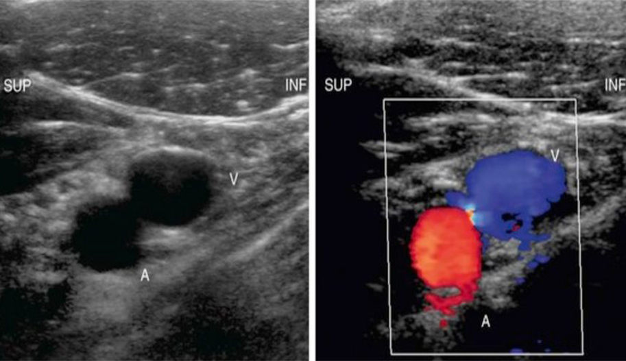
The axillary vein is located medial and inferior to the axillary artery. The subclavian vein lies anterior and inferior to the subclavian artery.
Risk Factors for Upper Extremity Deep Vein Thrombosis
Upper extremity deep vein thrombosis (DVT) can be classified as primary or secondary thrombosis based on its pathogenesis.
While the exact prevalence of symptomatic upper extremity DVT in the general population is unknown, it is estimated to be around 0.2%. About 90% of patients with DVT have lower extremity involvement, while the remaining 10% have upper extremity involvement.
The incidence of upper extremity DVT has increased with the rising use of central venous catheters. Earlier studies documented thrombosis in 2% to 12% of patients with central venous catheters. More recent studies report that 50% to 60% of patients with central venous catheters develop upper extremity DVT. The strongest independent predictor of upper extremity DVT in these patients was the presence of a catheter. The position of the catheter tip was found to be associated with the incidence of upper extremity DVT. Catheters located at the junction of the right atrium and superior vena cava and those within the mid-superior vena cava had the lowest incidence of thrombosis, while those with catheter tips in the brachiocephalic vein had a higher incidence. The ideal position for the catheter tip is at the cavoatrial junction. The material and diameter of the catheter were also found to affect the incidence of thrombosis. In the pediatric population, two-thirds of DVT cases occur in the upper extremity, unlike in adults, and are often secondary to catheter placement.
Cancer is a significant risk factor for upper extremity DVT due to changes in clotting factors, low-grade disseminated intravascular coagulation, and tumor-related stasis. Bilateral upper extremity DVT is more commonly found in patients with malignancy. The risk of thrombosis is significantly increased in patients with both cancer and central venous catheters.
Hypercoagulability (e.g., deficiencies in antithrombin, protein C, and protein S; presence of antiphospholipid antibodies) has been commonly found in idiopathic upper extremity DVT, where there is no clear associated illness or triggering factor. Recent studies have found coagulation abnormalities in 42% to 56% of patients with idiopathic upper extremity DVT. Temporary causes of hypercoagulability, such as estrogen use, pregnancy, and ovarian hyperstimulation, have been observed in women with idiopathic upper extremity DVT.
Other predisposing factors for upper extremity DVT include venous stasis, trauma, surgery, sepsis, and thoracic outlet obstruction secondary to anatomical anomalies. Interestingly, traditional risk factors associated with lower extremity DVT, such as obesity, advanced age, and surgery, were not significant risk factors for patients with catheter-unrelated upper extremity DVT. Patients with upper extremity DVT were more often male, younger, thinner, and more likely to be smokers compared to those with lower extremity DVT. Recent immobility and prior venous thromboembolism played a lesser role in patients with upper extremity DVT, although cancer was more prevalent.
Complications of Upper Extremity Deep Vein Thrombosis
The most serious complication of upper extremity DVT is pulmonary embolism. Once considered rare, the prevalence of pulmonary embolism in patients with upper extremity DVT is now reported to be between 7% and 36%. Clinically, patients with upper extremity DVT have been found to have a fourfold lower prevalence of symptomatic pulmonary embolism at presentation compared to those with lower extremity DVT.
Other, less common complications of upper extremity DVT include post-thrombotic venous insufficiency, loss of vascular access, superior vena cava syndrome, septic thrombophlebitis, and, rarely, venous gangrene.
Clinical Findings of Upper Extremity Deep Vein Thrombosis
The most common clinical presentation of upper extremity DVT is swelling and pain in the upper extremity and face. Edema typically does not pit. Less common signs and symptoms include skin discoloration, a sensation of coldness in the hand and forearm, tenderness in the affected vein, paresthesia, and numbness. Central venous catheter blockage can also be an indicator of thrombosis. Collateral veins may develop over the shoulder and chest wall, and a tender cord may be palpable, especially in the axillary region. However, these signs and symptoms are not specific, and objective testing is required to confirm the diagnosis. Additionally, many cases of upper extremity DVT, particularly those related to catheter placement, are asymptomatic.

How is Upper Extremity Venous Doppler Performed?
Compression technique with upper extremity venous color Doppler ultrasonography has become the preferred imaging method for diagnosing upper extremity deep vein thrombosis and is highly accurate for making this diagnosis. Sonography has the advantages of being non-invasive, requiring no radiation or contrast agents. It is a potentially portable examination and can be performed at the bedside in critically ill patients. It can be performed regardless of kidney function, and serial follow-up examinations can be done easily. Unlike venography, internal jugular and peripheral brachiocephalic veins can be evaluated even in the presence of thrombus in more peripheral veins. The limitations of color Doppler sonography include imaging the superior vena cava and the more central portions of the brachiocephalic veins. Additionally, small non-obstructive thrombi may go undetected in the subclavian vein due to the inability to compress this vessel beneath the clavicle. In patients with chronic deep vein thrombosis, it may also be challenging to distinguish a large collateral vessel from the native vein.
The
reported sensitivity of upper extremity color Doppler sonography for diagnosing upper extremity deep vein thrombosis ranges from 78% to 100%, and specificity ranges from 82% to 100%. False-positive examination results are considered rare. False negatives can occur due to limitations in the ability to compress veins.
There are differences in the recommended techniques and protocols for ultrasound examination of the upper extremity venous system. At our institution, in addition to imaging the internal jugular, subclavian, axillary, and brachial veins, the superficial basilic and cephalic veins are examined up to the level of the antecubital fossa.
The patient is positioned supine with the arm extended but without hyperabduction. The neck is slightly turned away from the side being examined. A compression technique is used starting at the internal jugular vein. Compression is always performed in the transverse plane because compression in the longitudinal plane can cause the probe to slip off the vein, potentially resulting in a false-negative result. All veins are compressed except in areas limited by the clavicle.