


Penile Doppler Ultrasound
Penile erection is a complex phenomenon coordinated by the interaction of arterial, venous, and nervous systems. A defect or lack of coordination in any of these systems can lead to erectile dysfunction, defined as the inability to achieve or maintain a sufficient penile erection for sexual intercourse. Erectile dysfunction can occur in young, middle-aged, and elderly men. It is strongly related to age and is estimated to affect approximately half of men between the ages of 40 and 70. This type of dysfunction can significantly impact the quality of life, affecting both physical and psychosocial well-being.
What is Penile Doppler?
There are many causes of erectile dysfunction. Penile color Doppler ultrasound is an imaging method used to investigate the cause of erection problems. The main purpose of the procedure is to measure the response of the penis to medications that increase blood flow to the penis.
The cause of erectile dysfunction can be organic (neurogenic, hormonal, vasculogenic, or drug-related), psychogenic, or mixed. The most common type of erectile dysfunction involves both psychogenic and organic components.
Why is Penile Doppler Ultrasound Performed?
Penile Doppler ultrasound is preferred for the initial evaluation of the penis because it can assess both the anatomy and dynamic blood flow; it is highly accessible, minimally invasive, and well tolerated by patients.
Color Doppler ultrasound provides the simultaneous visualization of blood flow while assessing the penile anatomy, plaques, fibrosis, tunica albuginea defects, masses, and fluid collections. It allows the evaluation of blood flow speed and direction.
The main role of imaging in patients with erectile dysfunction is to differentiate between vascular and non-vascular causes.
Penile Doppler ultrasound is primarily used to evaluate the integrity of the vascular mechanism and to rule out underlying arterial or venous insufficiency in patients who do not respond to or poorly respond to oral therapy. Erectile dysfunction is a common complication after pelvic surgery (mainly related to prostate, rectal, or bladder cancers). Therefore, penile Doppler ultrasound can be performed to confirm organic erectile dysfunction before penile prosthesis surgery.
Penile Doppler ultrasound is also recommended for the evaluation of erectile dysfunction after trauma, penile fibrosis, penile curvature (Peyronie’s disease), and candidates for penile implants.
How is Penile Doppler Ultrasound Performed?
The procedure is performed with the patient lying supine on the examination table. First, the entire penis is examined to rule out structural abnormalities. The diameter and blood flow velocities of the vessels responsible for erection on both sides of the penis are measured. Then, an injection (using a fine insulin needle called the penile Doppler needle) is administered, which induces an erection by increasing blood flow to the penis.
The procedure is generally painless. During the injection, patients may feel a slight and brief discomfort. Most patients report that their fear of the needle was unfounded. Erection begins within minutes after the injection, and blood flow velocities and patterns are recorded from both sides of the penis at 5, 10, 15, and 20-minute intervals.
Penile Doppler Ultrasound Results
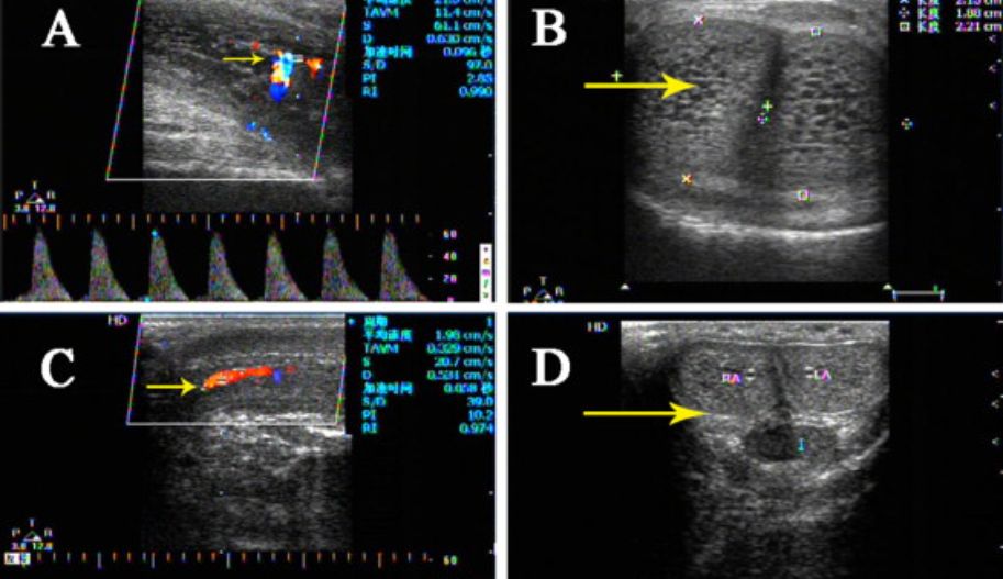
Commonly used hemodynamic parameters include peak systolic velocity (PSV), end-diastolic velocity (EDV), and resistance index (RI).
Through the evaluation of Doppler ultrasound parameters, the vascular response can be classified as normal or pathological (indicating arterial or venous insufficiency). A PSV of 35 cm/s or greater indicates arterial sufficiency. For venous sufficiency, EDV below 5 cm/s and RI greater than 0.8 are required.
Are There Any Risks with Penile Doppler Ultrasound?
Penile Doppler ultrasound is not a harmful procedure. Normally, erection returns to normal within half an hour to an hour after the procedure. Approximately 1% of patients may experience prolonged erection (priapism), where the penis does not return to its normal state. If such a condition, characterized by painful erection lasting up to 4 hours, occurs, a small needle intervention can solve the problem, and the penis returns to normal.
Peyronie’s Disease
Peyronie’s disease is a chronic benign fibrotic change of the penis, typically characterized by the development of fibrous plaques or nodules within the tunica albuginea, usually on the dorsal side. It can eventually cause penile deformity and is the most common cause of painful penile erection. Peyronie’s disease can be associated with other fibrotic conditions (e.g., Dupuytren’s contracture and Ledderhose disease) as well as vascular erectile dysfunction, including both arterial and venous insufficiency.
Penile Fracture
Penile fracture usually results from trauma to an erect penis, most commonly during sexual intercourse, leading to a tear in the tunica albuginea. Acutely, patients typically present with a history of a cracking sensation, pain, and sudden swelling of the penis accompanied by loss of erection.
Penile trauma can lead to long-term complications such as localized fibrous plaque formation, corpus scarring, and vascular erectile dysfunction.
Penile Doppler Ultrasound Prices 2026
Penile Doppler ultrasound prices in Istanbul vary by region and can differ even between institutions within the same area. For more information on Penile Doppler ultrasound prices, please contact us.