


What is the Uterine Film HSG?
Uterine film HSG is a radiological evaluation of the uterus and fallopian tubes and is mostly used for the evaluation of infertility. Other indications for HSG include evaluation of women with a history of recurrent spontaneous abortion, postoperative evaluation of women who underwent tubal ligation or opened tubal ligation, and evaluation of patients before myom Dectomy. The primary role of HSG is the evaluation of the fallopian tubes. Ultrasound is used to evaluate the endometrium ( such as abnormal uterine bleeding, polyps) and pregnancy, while magnetic resonance imaging (MRI) is more commonly used to evaluate the myometrium ( such as uterine contour, fibroids ) and ovaries. The number of HSGS has increased significantly over the past few years. This increase may most likely be due to (a) advances in reproductive medicine that have resulted in more successful in vitro fertilization procedures; (b)an increased trend towards women postponing pregnancy until later periods.
Hysterosalpingography ( HSG ) ( medicated uterine film ) is a very important imaging method in the investigation of pregnancy potential. In general, HSG allows the evaluation of the internal structure of the uterus and tubes, the opening of the tubes, thanks to X-rays by injecting a uterine film and a contrast agent into the tubes.
The liquid given in a normal woman fills the inside of the uterus without encountering any difficulties and passes into the tubes. From here it also spreads to the abdominal cavity.
What are the uses of uterine film HSG?
Hysterosalpingography is used to examine patients who need to be treated for infertility (infertility). It allows the radiologist to assess the shape and structure of the uterus, whether the fallopian tubes are open or closed.
Thanks to this examination, a blockage that prevents the union of sperm and eggs, or a uterine anomaly that prevents the fertilized egg from settling in the uterus, can be easily detected.
Uterine film is a valuable technique in the evaluation of infertile patients. Over the past decade, the number of women undergoing an infertility assessment has increased significantly. Uterine film HSG is considered a screening procedure for the study of infertility. Despite the development of other diagnostic tools such as MRI imaging, hysteroscopy and laparoscopy, hysterosalpingography is the main study in the investigation of the condition of the fallopian tubes. It has been reported that the uterine film has a high sensitivity, but low specificity, especially in the diagnosis of uterine cavity abnormalities. The technical quality of uterine film HSG is important to limit the factors that lead to misinterpretations.
When should the Medicated Uterine Film HSG be performed?
No specific patient preparation is required for HSG. There are two contraindications for HSG: pregnancy and active pelvic infection. The examination is performed on the 7th-12th of the menstrual cycle. it should be planned on days (1. the day is the first day of menstruation). Since the endometrium is thin during this proliferative phase, we both facilitate image interpretation and make sure that there is no pregnancy. In order to avoid a potential pregnancy, the patient should be told to refrain from sexual intercourse from the time of menstrual bleeding. If the patient has an irregular menstrual cycle or there is a possibility of pregnancy, it will be appropriate to perform the procedure after evaluating the serum β – HCG level and making sure that there is no pregnancy.
How to make a Painless Uterine HSG Film?
The patient lies on his back on the X-ray table and gathers his legs towards himself. First of all, the examination speculum is inserted and the cervix is seen. The vagina and cervix are wiped with a germ-killing solution. Then, a soft thin catheter (thin tube) with a balloon tip is inserted into the cervical canal. When the uterine film is taken, a drug containing fluid that appears on the X-ray film is used, it is gradually delivered into the uterus through a cannula. Fluoroscopic images are taken intermittently to evaluate the uterus and fallopian tubes.
At least four spot radiographs should be taken
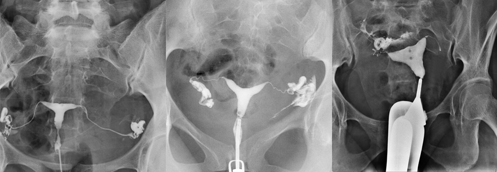
The first image ( Figure 1a) is obtained during early filling of the uterus and is used to evaluate any filling defects or contour abnormalities. Small filling defects are best seen at this stage. The second image (Figure 1b) is taken when the uterine cavity is completely filled. Although small filling defects can be hidden when the uterus is well opacified, it is at this stage that the shape of the uterus is best evaluated. The third image (Image 1c) is obtained to show and evaluate the fallopian tubes. The fourth image (Figure 1d ) should show the free intraperitonal spread of the contrast agent. Additional spot radiographs may be taken to detect any abnormalities observed. Oct. Oblique images can be taken when necessary to better view the tubes and prevent overlaps. At the end of the study, if the balloon closes the area where it was placed at the first placement, a radiography is obtained after deflating the balloon to evaluate the uterine lower segment. We do not use the tenaculum; if it is necessary to pull the uterus, we do this by gently pulling down the HSG catheter.
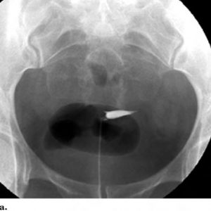
Image 1a. Spot radiography obtained at the early filling stage of the uterus. Small filling defects are best seen at this stage.
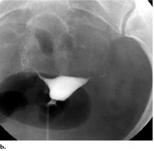
Image 1b In a radiograph obtained after the uterus is completely filled with a contrast agent, the initial parts of both fallopian tubes become opaque. Like images obtained at the early filling stage of the uterus, images obtained at full uterine distension allow the evaluation of defects and contour abnormalities. However, small filling defects may be hidden when the uterus is completely full.
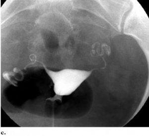
Figure 1c clearly shows the interstitial, isthmic and ampullary parts of both fallopian tubes.
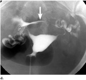
Figure 1d It is seen that the contrast agent is poured from the fallopian tubes into the intraperitoneal area. In this case, the shedding shows the outward curve of the uterine fundus(arrow).
Is Medicated Uterine Film HSG a painful procedure ?
Due to the fact that the cervix is held and pulled with an instrument called the tenaculum in a medicated uterine film made with old techniques, patients are afraid to have this procedure performed and / or request anesthesia, as it may cause pain.
Thanks to newly developed techniques, uterine film can be made painlessly. In addition to the fact that the devices and apparatuses used here are new technologies, the experience and experience of the team that makes the application are also very important.
What are the other advantages of Medicated Uterine Film HSG?
One of the most important advantages is the therapeutic property of HSG. After HSG withdrawal, small adhesions inside the tubes are opened.
If other parameters are normal in an infertile woman (with infertility problems), and pregnancy rates increase in the 6 months after HSG shooting.
For this reason, medicated uterine film HSG is an important method for both diagnosis and treatment in the treatment of infertility.
Womb Film HSG Prices 2025
By calling local health institutions, medical centers or hospitals directly, you can find out the prices of medical procedures such as uterine film or hysterosalpingography (HSG). You should also make sure that some health insurance may cover certain medical procedures.