


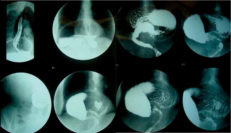
What Is A Stomach X-ray (Esophagus, Stomach, Duodenum X-ray)?
A Stomach and Duodenum X-ray; Diagnostic tests for evaluating the esophagus involve showing structural abnormalities, identifying functional anomalies, and measuring the esophagus’s exposure to stomach acid. The first radiological test of choice to assess the morphology and motor functions of the esophagus is a barium stomach X-ray.
The EGD (Esophagus, Stomach, Duodenum) X-ray is a procedure in which the doctor uses real-time X-rays called fluoroscopy and a liquid contrast material called barium to visualize your upper GI (gastrointestinal) tract. Barium will make your upper GI tract (esophagus, stomach, and the first part of the small intestine or duodenum) more visible on an X-ray. Fluoroscopy (real-time X-ray) makes it possible to see internal organs in motion. When the upper GI tract is coated with barium, the radiologist can see and assess the anatomy and function of the esophagus, stomach, and duodenum.
Difficulty swallowing, burning sensation in the throat, painful swallowing, chest pain, globus sensation, and pre-and post-treatment evaluation are the main indications for a barium stomach X-ray. Combined fluoroscopic evaluation during a barium stomach X-ray is more effective in diagnosing these conditions. When an esophageal disease is suspected, the first test for patients should be a barium stomach X-ray, including a full evaluation of the stomach and duodenum.
Preparation is required before the stomach X-ray. The patient should not eat, drink, or smoke for at least 8 hours before the procedure.
How Is An EGD X-ray Performed?
The test begins by having the patient drink a barium liquid that looks like plaster on an empty stomach. The swallowing function is evaluated through images taken during swallowing. Once the barium fills the stomach, the patient is turned upside down, and as images continue to be taken, the full contours of the fundus region are visible, and it can be determined if there is leakage into the esophagus, indicating reflux.
The barium is ingested quickly to fill the entire esophageal lumen. Using this method, cancer, large ulcers, and webs can be identified, and the contour, motility, and expansion ability of the esophagus can be effectively evaluated.
Continuous dynamic imaging is preferred for assessing pharyngeal and esophageal motility. In a normal adult, bolus passage through the 20-24 cm long esophagus is completed within 6-8 seconds. Retention and delayed emptying of barium in patients with esophageal motility disorders are significant findings.
After reaching the stomach, the folds of the stomach and ulcers in the stomach wall appear as outward bulging filling defects. Filling defects that protrude inward indicate the presence of a mass, suggesting stomach cancer. Small protruding filling defects on top of these filling defects indicate the presence of an ulcer that has developed on a tumor in the stomach.
In cases where the filling defects protrude outward directly from the stomach wall, benign ulcers caused by excess stomach acid are considered, while the filling defects on top of inward filling defects suggest gastric cancer ulcers.
The barium passes smoothly through the stomach outlet, and after passing, filling defects or persistent filling areas in the bulb and duodenum may indicate ulcers. Strictures caused by ulcers in the bulb and duodenum are also easily visible on this X-ray.
Why Is An EGD X-ray Performed?
An EGD X-ray may be performed by your doctor to investigate the cause of the following:
Nausea and vomiting, chest and abdominal pain, difficulty swallowing, unexplained weight loss, reflux (the return of partially digested food and digestive juices), unexplained vomiting, severe indigestion, blood in the stool (indicating internal GI bleeding),
The EGD X-ray can also detect the following:
Abnormal growths such as cancer, inflammation of the esophagus, stomach, and duodenum, abnormalities in the bowel wall, anatomical problems like intestinal malrotation (twisting of the bowel), esophageal varices, gastroesophageal reflux, hiatal hernia, scar tissue or strictures, blockages, and ulcers.
How Long Does An EGD X-ray Take?
The EGD X-ray procedure typically takes about 2 hours. If the barium moves slowly through your small intestine, the EGD X-ray procedure can take up to 5 hours.
EGD X-ray and Endoscopy
Endoscopy is a procedure that allows your doctor to look inside your body using an instrument called an endoscope. The endoscope has a small camera attached to a long, thin tube. The doctor passes it through a body passage or opening to view the inside of an organ. Endoscopes are sometimes used in surgeries, such as removing polyps from the colon.
EGD X-ray and Barium Esophagram
An EGD X-ray allows your doctor to see your esophagus, stomach, and the first part of the small intestine or duodenum. In contrast, a barium swallow examines the pharynx, esophagus, and the upper part of your stomach.
Preparation For An EGD X-ray
Preparation for an EGD X-ray requires fasting for 8 hours. You should discuss any medical conditions you have and all prescription and over-the-counter medications, vitamins, and supplements you take with your doctor.
To see your upper GI system clearly, your doctor will likely ask you not to eat, drink, smoke, or chew gum for 8 hours before the EGD X-ray.
Doctors do not recommend X-rays for pregnant women because X-rays can harm the fetus. If you are pregnant or think you might be, inform your doctor. Your doctor may suggest a different procedure.
If the benefits of the procedure outweigh the relatively small risk from the X-ray, the doctor may recommend an EGD X-ray for your child. Speak to your child’s doctor about the safety measures used to reduce your child’s exposure to X-rays during the procedure.
EGD X-ray Procedure
An EGD X-ray can be performed at a hospital or outpatient center. No anesthesia is required. The EGD X-ray procedure typically takes about 2 hours. If the barium moves slowly through your small intestine, the procedure may take up to 5 hours.
You may receive an injection of medication to slow down muscle movement in the small intestine. This makes it easier to see the structures of your organs on the X-rays.
Before the X-ray, you will need to drink 16 to 20 ounces (480 to 600 milliliters) of a milkshake-like drink. The drink contains barium, a substance that shows up well on X-rays.
For the EGD X-ray procedure, you will be asked to stand or sit in front of an X-ray machine and drink the barium, which will coat the inside surface of your upper GI tract. You will then lie on the X-ray table, and the technician will monitor the passage of the barium through your GI tract using fluoroscopy. The technician may press on your abdomen or ask you to change positions several times to ensure the barium coats your upper GI tract evenly.
At times, other forms of contrast, usually containing iodine, may be given to some patients orally. These alternative contrast materials may be used if the patient has recently had surgery in the GI tract or is allergic to other contrast agents. The radiologist will determine which type of contrast to use.
Children typically drink the barium contrast without resistance. If the child refuses to drink the contrast, the radiologist may need to pass a small tube into the stomach to complete the examination.
After The EGD X-ray
You may experience cramping and bloating in your abdomen for the first hour after the procedure.
After leaving the hospital or outpatient center, you can resume most normal activities.
For several days, your stools may be white or light-colored due to the barium remaining in your GI tract.
Instructions will explain how to clear the remaining barium from your GI tract. Be sure to follow all instructions.
EGD X-ray Results
Abnormal results in the esophagus may indicate the following conditions:
Achalasia, diverticulum, esophageal cancer, esophageal stricture (benign), hiatal hernia, ulcers.
Abnormal results in the stomach may indicate the following conditions:
Stomach cancer, benign gastric ulcer, gastritis, polyps (usually non-cancerous tumors that grow in the mucous membrane), pyloric stenosis (narrowing).
Abnormal results in the small intestine may indicate the following conditions:
Malabsorption syndrome, inflammation, and irritation of the small intestine, tumors, ulcers.
The EGD X-ray test may also be performed for the following conditions:
Annular pancreas, duodenal ulcer, gastroesophageal reflux disease, gastroparesis, intestinal obstruction, lower esophageal ring, primary or idiopathic intestinal pseudo-obstruction.
EGD X-ray Prices 2026
EGD X-ray prices in Istanbul vary from region to region and even between institutions in the same area. For information on EGD X-ray prices, you can call 02126321059.